
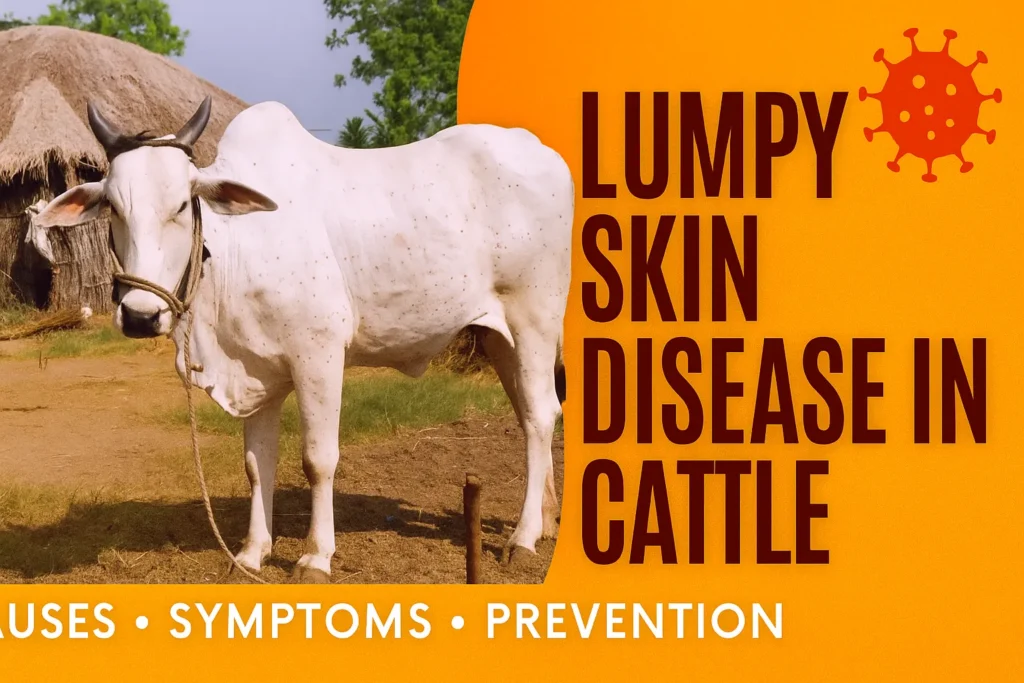
Lumpy Skin Disease: Symptoms, Treatment, and Prevention in Cattle
What is Lumpy Skin Disease?
Lumpy Skin Disease (Lumpy Skin Disease) is a viral infectious disease that primarily affects animals. It is caused by the Lumpy Skin Disease Virus (LSDV), which is a member of the Poxviridae family, Chordopoxvirinae subfamily, and Capripoxvirus genus. Capripoxvirus is also known as Neethling virus. Lumpy Skin Disease is an acute disease that affects animals for 3 to 15 days.
Lumpy Skin Disease in Cattle : THE RAJASTHAN EXPRESS
| Disease Classification |
|
|---|---|
| Etiological Agent |
|
| Host Range & Susceptibility |
|
| Epidemiology & Geographic Distribution |
|
| Transmission Dynamics |
|
| Clinical Presentation |
|
| Diagnostic Modalities |
|
| Vaccination & Control Strategies |
|
| Therapeutics & Supportive Care |
|
| Economic & Public Health Considerations |
|
| Comprehensive Symptoms of Lumpy Skin Disease: THE RAJASTHAN EXPRESS | |
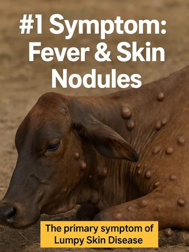
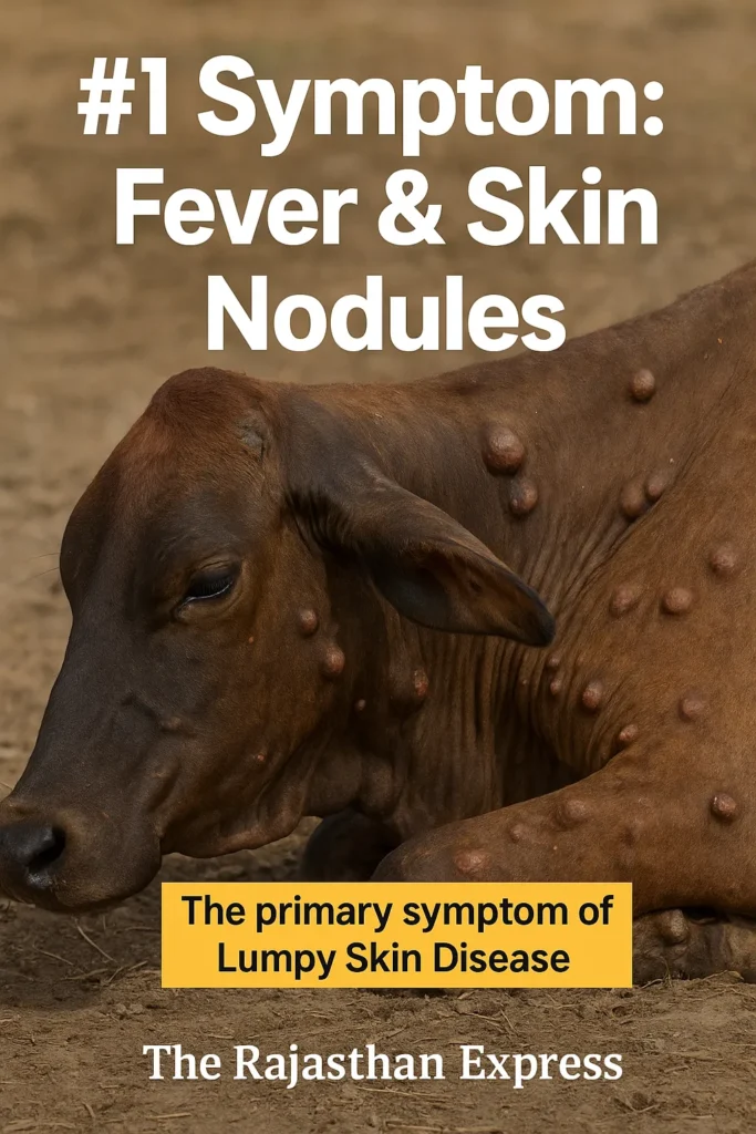
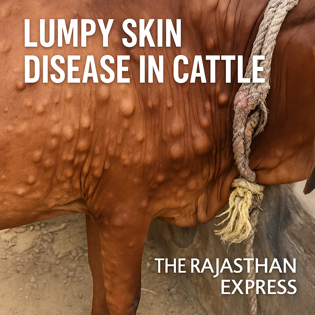
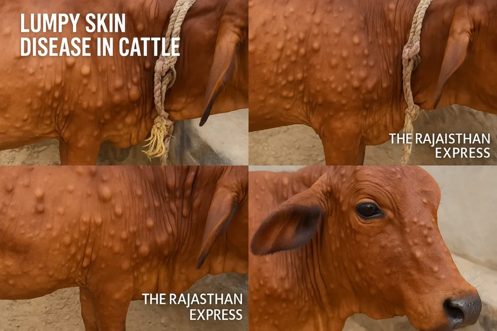
Symptoms of Lumpy Skin Disease in Cattle
This is a cattle disease in which symptoms such as fever, nodules on the skin/mucous membranes/internal organs, physical weakness, swollen lymph nodes, skin inflammation (edema), and sometimes death are seen. The emergence of nodules on the animal’s skin is the main identification of this disease, which causes the animal to suffer from severe inflammation. Also, high fever, inflammation in the respiratory tract, and lameness in the legs are observed in the animal.
Outbreak History in India
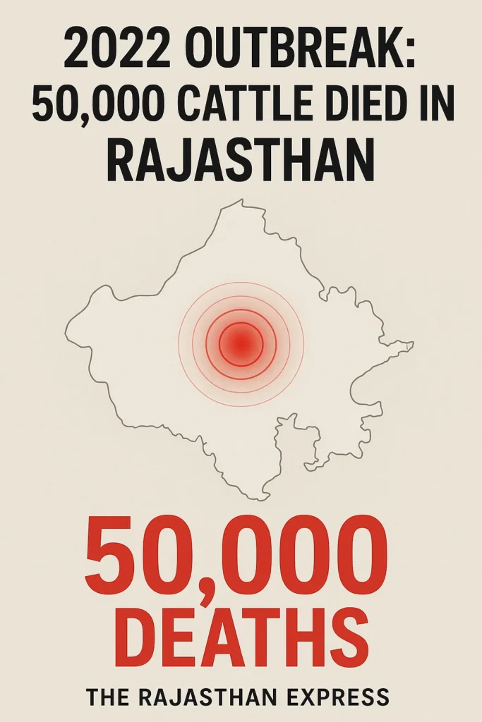
In India, this disease was first recorded in the state of Odisha. Lumpy Skin Disease (LSD) affected more than 400,000 cattle in Rajasthan, where the mortality rate was quite high. In Rajasthan alone, about 50,000 cows died, which indicates the fatality of this disease. For this reason, the World Organisation for Animal Health (WOAH) declared it a disease of the Epizootic category. Epizootic diseases affect a large number of animals in a large area. In addition to Lumpy Skin Disease, diseases like Foot-and-Mouth Disease (FMD) and Rinderpest also fall under this category.
Lumpy Skin Disease Outbreak in Europe
The World Organisation for Animal Health (WOAH) has acknowledged the official notifications and follow-up reports concerning two recent outbreaks of Lumpy Skin Disease (LSD) in France and Italy. These represent the first confirmed cases of lumpy skin disease in Europe in these countries, highlighting an urgent need for veterinary oversight and monitoring to prevent further spread. Both France and Italy have already begun taking action in line with international disease control standards.
Read More About LSD Outbreak in Europe : Lumpy Skin Disease Outbreak in Europe
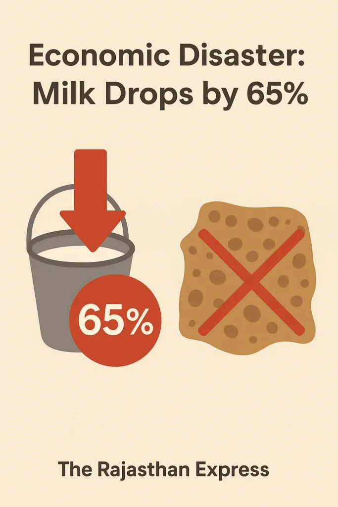
Economic Impact on Livestock Farmers
Lumpy Skin Disease, like Mastitis, causes heavy economic losses to farmers and cattle rearers. As seen in 2022 in Rajasthan, India, where the death of about 50,000 cows caused severe economic losses to local cattle rearers. This disease can cause a temporary reduction in milk production, temporary/permanent infertility in bulls, skin damage (which reduces the value of leather), and death. Exotic cows of the Bos taurus species are more susceptible to this disease compared to Bos indicus, i.e., Indian indigenous breeds. Asian buffalo (Bubalus spp.) can also be affected by this disease.
Clinical Signs and Disease Progression
In acutely infected animals, in the initial stage of the disease, a high fever (pyrexia) of up to 41°C is observed, which can last for a week (normal cow temperature is 38°C). All superficial lymph nodes (lymph glands) become swollen. In milch cows, milk production decreases. Within 7–19 days of virus infection (acute stage), lesions develop on the body, especially on the head, neck, udder, testicles, vagina, and anal area.
Characteristics of Skin Lesions
- Numerous, with clear boundaries or attached
- Hard, flat-surfaced papules/nodules of 0.5–5 cm diameter
- These develop in the upper (epidermis) and middle layers (dermis) of the skin and can sometimes spread to the underlying muscles.
- On cutting, a creamy-gray or white substance comes out, which may initially ooze serum.
- Within 2 weeks, a cone-shaped dead tissue (necrotic plug or “sit-fast”) may form in them.
Various Capri pox virus strains are responsible for this disease. These are antigenically similar to Sheep pox and Goat pox viruses but differ at the genomic level.
Transmission and Vaccine for Lumpy Skin Disease
LSDV transmission mainly occurs through insect vectors (arthropods), while insect-free direct contact transmission is less effective. An important fact is that it is not a zoonotic disease, meaning it does not spread from animals to humans. Vaccines developed from weakened live viral strains of cattle, sheep, and goats are available against this virus. In India, initially, the Goat Pox Vaccine was found to be less effective. Later, two major Indian government institutions—the National Research Centre on Equines (NRCE) and the Indian Veterinary Research Institute (IVRI)—jointly developed the “Lumpi-ProVacInd” vaccine, which yielded excellent results. This is the first indigenous homologous vaccine for LSD, made from the Ranchi strain of Capri pox virus.
Etiology of Lumpy Skin Disease
Lumpy skin disease is mainly caused by Capripoxvirus of the Poxviridae family, which is also known as Neethling virus. LSDV is a double-stranded DNA virus. Additionally, this disease can also be caused by Sheep pox and Goat pox viruses. Like other members of the Poxviridae family, Capripoxviruses are brick-shaped. This virus is one of the largest in size, with an average size of 320 nm × 260 nm. It is a highly contagious disease that can spread from one animal to another.
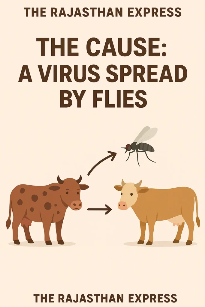
Modes of Transmission of Lumpy Skin Disease
- In animals infected with Lumpy Skin Disease, the virus can survive in saliva for 11 days, in semen for 22 days, and in skin nodules for up to 33 days.
- This disease spreads more during seasons with high temperature and humidity, especially during summer and monsoon.
- The main spread of Lumpy Skin Disease occurs through blood-sucking insects such as mosquitoes and flies.
- This disease can also spread through contact with the blood, nasal discharge, tears (lacrimal secretion), semen, and saliva of infected animals.
- It can also spread in calves, especially during suckling.
- Transmission of the virus through semen is also possible, meaning it can spread during mating, although this is very rare.
- One case of transmission through the uterus has been recorded.
Epidemiology of Lumpy Skin Disease
- Primary Hosts and Susceptibility
- Lumpy Skin Disease primarily affects cows and buffaloes (Bos indicus, Bos taurus, Bubalus bubalis).
- Holstein Friesian breed has been found to be more susceptible to this disease.
- Native Indian breeds have shown relatively higher resistance to Lumpy Skin Disease.
2. Other Species and Zoonotic Status
- Infection cases have also been recorded in some wildlife species such as springbok, eland, oryx, gaur, and banteng.
- This virus is not infectious to humans, meaning it is not a zoonotic disease.
- No confirmed cases of Lumpy Skin Disease (LSD) infection in sheep-goats have been reported so far.
3. Morbidity and Mortality Rates
Morbidity (Spread rate):
- Typically 10–20%, occasionally reaching up to 45%.
- In 2022 in India, the spread rate of this disease was extremely high, causing approximately 4–5 lakh cows to be infected in Rajasthan state alone.
Mortality (Death rate):
- Usually between 1–5%.
- However, in 2022 in Rajasthan, its impact was very severe, during which over 50,000 cows died.
4. Source of the Virus
- Capripox Virus is found in skin nodules, scabs, blood, saliva, eye-nasal secretions, and semen of infected animals.
- The virus can survive in blood for 7–21 days.
5. Environmental Sensitivity
Temperature:
- Virus can be destroyed at 55°C in 2 hours and at 65°C in 30 minutes
- At -80°C temperature, it can survive for 10 years.
- In pus or fluid present in infected tissues, this virus can survive at 4°C for 6 months.
pH level:
- This virus is sensitive to acidic and alkaline pH.
- Between pH 6.6–8.6, at 37°C, the virus can remain effective for 5 days.
Environmental persistence:
- LSDV or Capripox Virus can survive in dried lesions for 33 days or longer.
- It can remain active for several months in dark, infected animal sheds.
Geographic Presence and History
- This disease is endemic in Africa and periodically causes outbreaks there.
- Since 2012, it has rapidly spread to the Middle East, Europe, and Asia.
- Several outbreaks have emerged in South Asia since 2019.
- The first case of Lumpy Skin Disease in India was recorded in 2019 in Odisha state.
- In 2022, the most severe outbreak of this disease was observed in Rajasthan, where very high animal mortality was recorded.
Capripox Virus: Chemical Sensitivity
This virus is sensitive to the following chemicals:
- Ether (20%)
- Chloroform
- Formalin (1%)
- Sodium dodecyl sulfate
- Phenol (2%, for 15 minutes)
- Sodium hypochlorite (2–3%)
- Iodine compounds (1:33)
- Virkon® (2%)
- Quaternary ammonium compounds (0.5%)
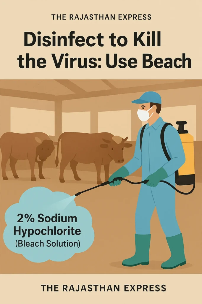
The most effective disinfectant for infection prevention is considered to be 2% sodium hypochlorite.
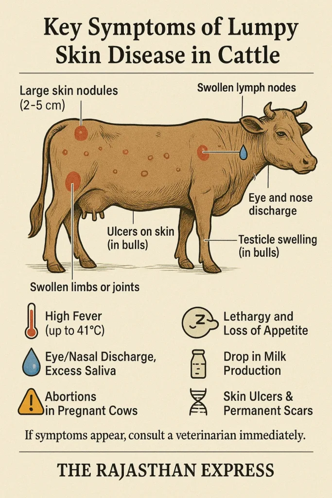
Comprehensive Symptoms of Lumpy Skin Disease
- Formation of Large Nodules:
Painful and unbearable nodules (approximately 2–5 cm in size) develop on the animal’s body. These nodules are the primary identifying sign of the disease. - General Symptoms:
- Lethargy, loss of appetite, and weakness
- Nasal discharge, watery eyes, and excessive salivation indicating infection severity
- Risk of abortion in pregnant cows
- High Fever:
High fever (approximately 41°C) may be observed about 4–14 days after infection, persisting for several days. - Swelling in Internal Organs:
- Swelling in respiratory system and lymph nodes
- Swollen lymph nodes, inflammation in airways, discharge from nose and eyes, fluid accumulation in legs, testicles or udders are common signs
- In male animals, scrotal swelling may temporarily affect spermatogenesis, reducing fertility
- Reduced Milk Production:
Milk-producing animals may experience significant decline in milk yield due to the disease. - Development of Ulcers:
Within 1–2 weeks of nodule formation, ulcers appear on various body parts (e.g., head, neck, thighs, genitals). - Persistent Skin Damage:
Even after treatment, permanent scars or marks remain on the skin, potentially reducing the animal’s commercial value. - Variation in Susceptibility:
- Indigenous breeds (e.g., Bos indicus) are less susceptible to this disease compared to exotic breeds (Bos taurus).
- Exotic breeds are more affected due to higher milk production, softer skin, and lower immunity.
- Calves and lactating cows may also be more affected due to weaker immunity.
Incubation Period and Disease Nature
- Incubation period is considered 4–14 days, with maximum observed cases up to 28 days.
- Lumpy Skin Disease is an acute infection, and due to its intense outbreak, it is also considered an epizootic disease.
Diagnosis and Management of Lumpy Skin Disease
- Based on Clinical Signs:
Preliminary diagnosis can be made based on symptoms like body nodules, fever, discharge, etc. - ELISA (Enzyme-Linked Immunosorbent Assay):
Confirms virus by measuring antibody levels.
Safe, reliable, and widely used test. - IPMA (Immunoperoxidase Monolayer Assay):
Identifies tissues infected with LSD virus. - Virus Neutralisation Test:
Confirms presence of specific disease-resistant antibodies.
Considered the “Gold Standard” test. - DIVA PCR (Differentiating Infected from Vaccinated Animals):
This test differentiates between vaccine-derived and natural virus.
Other Useful Tests
- Electron microscopy
- Immunohistochemistry
- Gene sequencing
- Western blot (highly accurate but expensive)
- Development of novel ELISA kits is underway.
Differential Diagnosis
Diseases with symptoms similar to LSD include:
- Bovine herpes mammillitis
- Papular stomatitis
- Pseudocowpox
- Cowpox
- Dermatophilosis
- Insect bite reactions
- Besnoitiosis
- Rinderpest (Cattle plague)
- Hypoderma bovis infection
- Photosensitization
Treatment of Lumpy Skin Disease
- No specific or complete treatment for LSD is available.
- In early disease stages, antibiotics, anti-inflammatory drugs, and analgesics may be used to prevent secondary infections.
- Supportive therapy (e.g., fluids, nutritious diet, ointments) is also used to relieve the animal and reduce disease severity.
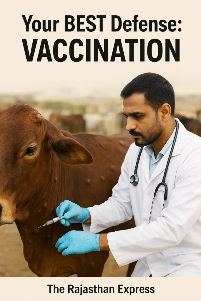
Vaccination for Lumpy Skin Disease
Primary Vaccine: Neethling Strain
- Considered the most effective vaccine for LSD.
- Administered to calves above 6 months of age.
- Provides immunity for approximately 3 years.
- Particularly effective in cows.
Alternative Vaccines
1. Goat Pox Vaccine (GTPV):
- Considered less effective, but results improve at higher doses.
- Initially used in India.
2. Lumpi-ProVacInd Vaccine (Indigenous Vaccine):
- Developed by NRCE (National Research Centre on Equines) and IVRI (Indian Veterinary Research Institute).
- Approval: CDSCO
- Launched: 2022–2023
- First homologous vaccine made from Capripox virus’s “Ranchi strain”.
- Currently the most widely used vaccine in India.
- Provides excellent protection and disease-resistant results.
Control Measures for Lumpy Skin Disease
1. Recommended Disinfectants
The following chemicals can be used to kill the LSD virus:
- Ether (20%)
- Chloroform
- Formalin (1%)
- Sodium Dodecyl Sulfate (Detergent)
- Sodium Hypochlorite (2%) (most widely used)
- Phenol (2%) – Minimum 15 minutes of contact required
2. Effective Control Strategies
- Isolate infected animals from healthy livestock.
- Properly dispose of dead animals (e.g., burial or incineration).
- Regularly clean and disinfect farms, equipment, and sheds.
- Spray Formalin (1%) or Sodium Hypochlorite (2%) weekly.
- Restrict animal movement in infected areas.
- Avoid feeding milk from infected cows to calves; separate calves from infected animals.
Post-Mortem Findings in Lumpy Skin Disease
Key Pathological Observations
- Skin Nodules:
- Painful nodules deep in the skin with visible hemorrhaging, vascular obstruction, and inflammation.
- Scabs on the surface and necrotic (dead tissue) cores within nodules.
- Mucous Membrane Afflictions:
- Ulcers and inflammation in the oral and nasal cavities.
- Swelling/lesions in the gastrointestinal tract, lungs, testicles, and bladder.
- Systemic Organ Involvement:
- Affects multiple organ systems.
- Infection spreads from skin to internal organs.
- Bronchopneumonia:
- Severe inflammation in lungs/respiratory tract (bronchi and alveoli).
- Major cause of respiratory distress and death.
- Lymph Node Enlargement:
- Marked swelling and hardening in superficial/deep lymph nodes.
- Indicates immune activation and localized infection.
- Synovial Involvement:
- Synovitis: Inflammation of joint membranes.
- Tenosynovitis: Inflammation of tendon sheaths.
- Fibrin deposits around joints causing lameness and stiffness.
Fever, skin nodules & reduced milk yield? Learn transmission via insects, prevention with vaccines, and supportive care for lumpy skin disease in cattle. Veterinary guidance included.
THE RAJASTHAN EXPRESS
Frequently Asked Questions About Lumpy Skin Disease (LSD)
What are the most effective vector control strategies for preventing lumpy skin disease transmission?
- Insecticide spraying: Use cypermethrin or deltamethrin on cattle and barns every 7-10 days
- Physical barriers: Install insect-proof netting in cattle housing
- Environmental management: Eliminate stagnant water and manure piles (breeding sites for mosquitoes and flies)
- Movement restrictions: Disinfect livestock transport vehicles before/after entry
- Seasonal vigilance: Intensify control during monsoon/summer when outbreaks peak
How do Neethling-based vaccines compare to sheep/goatpox vaccines for LSD prevention?
Comparison Summary:
- Neethling vaccine provides 85–95% protection and immunity for up to 3 years.
- Sheep/goatpox vaccine offers 60–75% protection and requires booster doses.
- Indian alternative “Lumpi-ProVacInd” (Ranchi strain) shows ≥90% efficacy.
What is the evidence-based supportive care protocol for LSD-infected cattle?
- Antibiotics: Oxytetracycline for 5-7 days to prevent secondary infections
- Anti-inflammatories: Flunixin meglumine (1.1-2.2 mg/kg) for fever/pain
- Wound care: Daily cleaning of nodules with povidone-iodine
- Hydration: Electrolyte supplements for dehydrated animals
- Isolation: Immediate separation from herd to reduce stress
Why are Bos taurus breeds more susceptible to severe LSD than Bos indicus?
- Skin thickness: Bos indicus have thicker skin that limits vector penetration
- Immune response: Bos taurus show delayed interferon production
- Outbreak data: Exotic breeds (e.g., Holstein) suffered disproportionately in Rajasthan 2022 outbreak
- Thermotolerance: Indigenous breeds better handle fever-related stress
What are the long-term economic impacts of LSD on dairy vs. beef operations?
- 20–65% milk yield reduction for 3–6 months
- 25–50% abortion risk in pregnant cows
- 60–80% hide devaluation from permanent scarring
- 15–25% weight loss delays market readiness
How does climate change influence LSD spread into non-endemic regions?
- Warmer temperatures: Extend vector activity seasons by 2–4 months
- Increased rainfall: Creates breeding sites for mosquitoes (secondary vectors)
- Case evidence: Rajasthan 2022 outbreak coincided with extreme monsoon conditions
- Projected risk: May reach Western Europe/US Midwest by 2030
Which diagnostic tests for LSD are most reliable at different disease stages?
- Early stage (1–7 days): PCR on blood/skin swabs – 98% accurate
- Acute stage (8–14 days): Virus isolation + histopathology – gold standard
- Recovery stage (>14 days): ELISA or Virus Neutralization – 90–95% accuracy
Can LSDV transmit via semen in artificial insemination, and for how long?
- Yes: Virus can persist in semen for up to 22 days post-infection
- Risk management:
- Test semen weekly with PCR during outbreaks
- Quarantine infected bulls for at least 22 days post-recovery
What are the management strategies for LSD in pregnant cows and calves?
- 25–50% abortion risk – isolate in stress-free zones
- Progesterone therapy may help maintain pregnancy
- 80% mortality in neonates – feed heat-treated colostrum (56°C for 60 min)
- Vaccinate calves at 3–6 months with half-dose Neethling vaccine
How long do LSD-recovered cattle remain contagious, and what are quarantine best practices?
- Contagious duration:
- Skin scabs: up to 33 days
- Blood: 7–21 days
- Quarantine best practices:
- Isolate infected/recovered animals for at least 33 days
- Disinfect barns with 2% sodium hypochlorite weekly
- Dispose carcasses by deep burial or incineration