 By
ByWhat Is Aspiration Pneumonia in Dogs? (Definition, Causes, and Scientific Explanation)
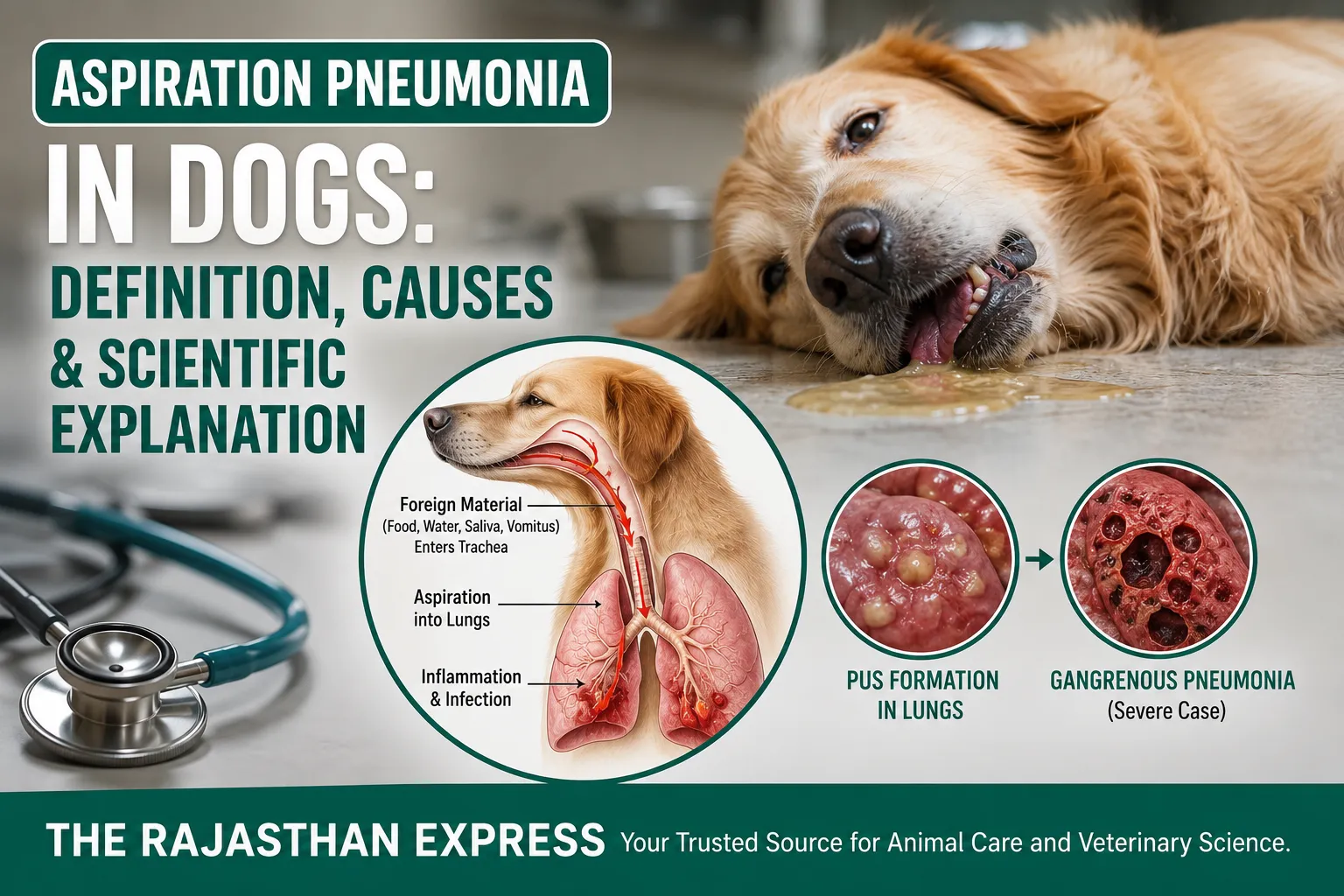
Aspiration Pneumonia is a serious infection (Infectious Pneumonia) or inflammation (Inflammatory Lung Disease) of the lungs that occurs when food, water, milk, medicine, vomitus, saliva, or any other foreign substance accidentally enters the trachea (windpipe) instead of the esophagus (food pipe) and reaches the lungs. This process is called Aspiration.
Under normal circumstances, during swallowing, the epiglottis closes the trachea, preventing food or liquid from entering the lungs. However, if this protective mechanism fails, foreign material can reach the alveoli and damage the lungs.
Once these substances reach the lungs, they cause acute inflammation in the alveoli, surrounding lung tissue, and the pleural membrane (pleura). If bacteria also enter along with the material, the infection can spread rapidly, leading to the formation of pus (suppuration) in the lungs.
In severe cases, impaired blood flow can cause necrosis (death) of lung tissue. If this necrosis becomes extensive and gangrene develops in the dead tissue, the condition is called Gangrenous Pneumonia. In such cases, lung abscesses, foul‑smelling pus, and sometimes spread of infection to the pleura causing pleuritis or pyothorax can also occur.
Note: Not every aspiration pneumonia is gangrenous pneumonia. Gangrenous pneumonia develops only in severe cases where there is extensive necrosis and gangrene of lung tissue.“`html
Aspiration Pneumonia in Dogs
| Medical Name |
Aspiration Pneumonia (AP) Bacterial Aspiration Pneumonia (BAP) Aspiration Pneumonitis (Chemical Pneumonitis) |
|---|---|
| Disease Type |
|
| Affected Species |
|
| Definition | Aspiration pneumonia is a potentially life-threatening lung disease that develops when food, milk, water, saliva, vomitus, gastric acid, medicines or other foreign material accidentally enters the trachea instead of the esophagus and reaches the lungs, resulting in chemical injury, bacterial infection, or both. |
| Primary Organ Affected | Lungs (Alveoli, Bronchi, Bronchioles, Pleura) |
| Normal Protective Mechanism |
|
| Pathogenesis | Foreign material enters the lungs → Chemical injury to alveoli → Surfactant destruction → Atelectasis → Inflammation (Chemical Pneumonitis) → Secondary bacterial multiplication → Aspiration Pneumonia → Necrosis → Lung Abscess → Gangrenous Pneumonia (severe cases). |
| Types |
|
| Common Aspirated Materials |
|
| Major Causes in Dogs |
|
| Major Causes in Large Animals |
|
| Major Risk Factors |
|
| Commonly Affected Breeds |
|
| Natural Lung Defense System |
|
| When Defense Fails |
|
| Chemical Injury | Gastric acid rapidly destroys pulmonary surfactant, causes alveolar collapse (atelectasis), severe inflammation, pulmonary edema and chemical pneumonitis (Mendelson’s Syndrome in humans). |
| Secondary Bacterial Infection | Following chemical injury, bacteria rapidly multiply inside damaged alveoli, producing bacterial aspiration pneumonia. |
| Lipid Pneumonia | Caused by aspiration of mineral oil or oil-based medicines leading to foamy macrophages and chronic granulomatous inflammation. |
| Important Cells |
|
| Main Bacteria (Dogs) |
Anaerobes
|
| Main Bacteria (Large Animals) |
|
| Clinical Signs |
|
| Emergency Signs |
|
| Diagnosis |
|
| Typical X-ray Findings | Bronchoalveolar pattern involving the cranial ventral lung lobes with alveolar consolidation. |
| Treatment |
|
| Supportive Care |
|
| Complications |
|
| Prognosis | Good when diagnosed and treated early. Guarded to poor in severe acid aspiration, gangrenous pneumonia, extensive necrosis, pyothorax or delayed treatment. |
| Prevention |
|
| Sources: Merck Veterinary Manual; Ettinger & Feldman’s Textbook of Veterinary Internal Medicine; BSAVA Manual of Canine and Feline Respiratory Medicine; Veterinary Clinics of North America: Small Animal Practice; Fossum’s Small Animal Surgery; Current Veterinary Therapy; The Rajasthan Express. | |
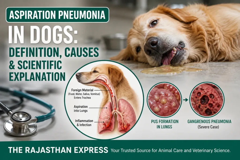
Scientific Facts – How does the lungs’ defense system work and when does it fail?
The lungs come into contact with millions of dust particles, bacteria, viruses, and other microorganisms daily. Yet, most of the time, infection does not occur because the respiratory system has several natural defense mechanisms.
- Mucociliary Clearance: The inner surface of the trachea and bronchi has microscopic hair‑like structures called cilia. A sticky mucus layer is present on top of them. Dust, bacteria, and other particles first get trapped in the mucus. Then, the cilia continuously move in one direction, pushing this mucus towards the pharynx (throat). From there, it is either expelled by coughing or swallowed, where gastric acid destroys most microorganisms.
- Alveolar Macrophages (Dust Cells): This is the second line of defense. Alveolar macrophages (dust cells) are present in the alveoli (air sacs of the lungs). If any bacteria, virus, or dust particle bypasses the mucociliary clearance and reaches the alveoli, these macrophages engulf and destroy it through phagocytosis. Simultaneously, they release cytokines and other chemicals to activate the immune system.
- Surfactant: This is a lubricant‑like substance lining the inside of the alveoli. Surfactant is a specialized lipoprotein substance present on the inner surface of the alveoli. Its main functions are:
- Preventing the alveoli from collapsing during exhalation.
- Maintaining lung compliance (the ability of the lungs to expand).
- The SP‑A and SP‑D (Surfactant Proteins) present in it help identify bacteria, viruses, and fungi and assist in activating immune cells.
- They enhance the pathogen‑destroying ability of alveolar macrophages.
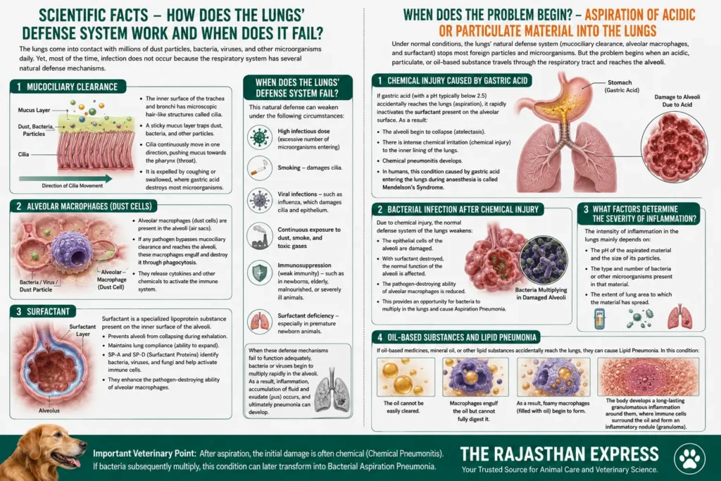
When does the lungs’ defense system fail?
Under the following circumstances, this natural defense can weaken:
- High infectious dose (excessive number of microorganisms entering)
- Smoking – damages cilia.
- Viral infections – such as influenza, which damages cilia and epithelium.
- Continuous exposure to dust, smoke, and toxic gases
- Immunosuppression (weak immunity) – such as in newborns, elderly, malnourished, or severely ill animals.
- Surfactant deficiency – especially in premature newborn animals.
When these defense mechanisms fail to function adequately, bacteria or viruses begin to multiply rapidly in the alveoli. As a result, inflammation, accumulation of fluid and exudate (pus) occurs, and ultimately pneumonia can develop.
When Does the Problem Begin? – Aspiration of Acidic or Particulate Material into the Lungs
Under normal conditions, the lungs’ natural defense system (mucociliary clearance, alveolar macrophages, and surfactant) stops most foreign particles and microorganisms. But the problem begins when an acidic, particulate, or oil‑based substance travels through the respiratory tract and reaches the alveoli.
1. Chemical injury caused by gastric acid
If gastric acid (with a pH typically below 2.5) accidentally reaches the lungs (aspiration), it rapidly inactivates the surfactant present on the alveolar surface. As a result:
- The alveoli begin to collapse (atelectasis) .
- There is intense chemical irritation (chemical injury) to the inner lining of the lungs.
- Chemical pneumonitis develops.
- In humans, this condition caused by gastric acid entering the lungs during anaesthesia is called Mendelson’s Syndrome.
2. Bacterial infection after chemical injury
Due to chemical injury, the normal defense system of the lungs weakens:
- The epithelial cells of the alveoli are damaged.
- With surfactant destroyed, the normal function of the alveoli is affected.
- The pathogen‑destroying ability of alveolar macrophages is reduced.
- This provides an opportunity for bacteria to multiply in the lungs and cause Aspiration Pneumonia.
3. What factors determine the severity of inflammation?
The intensity of inflammation in the lungs mainly depends on:
- The pH of the aspirated material and the size of its particles.
- The type and number of bacteria or other microorganisms present in that material.
- The extent of lung area to which the material has spread.
4. Oil‑based substances and Lipid Pneumonia
If oil‑based medicines, mineral oil, or other lipid substances accidentally reach the lungs, they can cause Lipid Pneumonia. In this condition:
- The oil cannot be easily cleared.
- Macrophages engulf the oil but cannot fully digest it.
- As a result, foamy macrophages (filled with oil) begin to form.
- The body develops a long‑lasting granulomatous inflammation around them, where immune cells surround the oil and form an inflammatory nodule (granuloma).
Important Veterinary Point: After aspiration, the initial damage is often chemical (Chemical Pneumonitis) . If bacteria subsequently multiply, this condition can later transform into Bacterial Aspiration Pneumonia.
Macrophage – The Body’s First Line of Cellular Defense : Role in Aspiration Pneumonia in Dogs
A macrophage is one of the largest white blood cells (WBCs) in our body. It is a specialized immune cell that develops from a cell called a monocyte. When a monocyte leaves the blood and enters a tissue, it enlarges and becomes a macrophage.
Its main job is phagocytosis. In this process, the macrophage engulfs any foreign and dangerous material – such as bacteria, viruses, fungi, parasites, dead cells, and other foreign particles – and then completely destroys them with the help of enzymes. This is exactly like a sanitation worker picking up and disposing of garbage.
But the macrophage does not just eat pathogens; it also gives further instructions to the body’s entire immune system. It performs two more very important functions: through antigen presentation, it activates T‑lymphocytes to initiate the body’s adaptive immunity. Additionally, it secretes cytokines and other chemical messengers to assist in inflammation, infection control, and tissue repair.
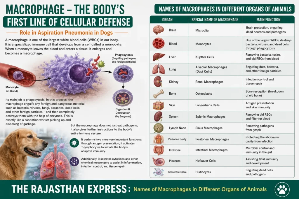
| Names of Macrophages in Different Organs of Animals | ||
| Organ | Special Name of Macrophage | Main Function |
|---|---|---|
| Brain | Microglia | Brain protection, engulfing dead neurons and pathogens |
| Blood | Monocytes | One of the largest WBCs; destroys bacteria, viruses, and dead cells through phagocytosis |
| Liver | Kupffer Cells | Removing bacteria, toxins, and old RBCs from blood |
| Lung | Alveolar Macrophages (Dust Cells) | Engulfing dust, bacteria, and other foreign particles |
| Kidney | Renal Macrophages | Infection control and tissue repair |
| Bone | Osteoclasts | Bone resorption (breakdown of old bone) |
| Skin | Langerhans Cells | Antigen presentation and skin immunity |
| Spleen | Splenic Macrophages | Removing old RBCs and filtering blood |
| Lymph Node | Sinus Macrophages | Removing pathogens from lymph |
| Peritoneal Cavity | Peritoneal Macrophages | Protecting the abdominal cavity from infection |
| Intestine | Intestinal Macrophages | Microbial control and immunity in the gut |
| Placenta | Hofbauer Cells | Assisting fetal immunity and development |
| Connective Tissue | Histiocytes | Engulfing dead cells and pathogens |
| The Rajasthan Express : Names of Macrophages in Different Organs of Animals | ||
Etiology of Aspiration Pneumonia: Causes of Aspiration Pneumonia In Dogs
Etiology of Aspiration Pneumonia in Dogs and Other Animals (Causes of Aspiration Pneumonia)
Aspiration pneumonia develops when food, liquid, saliva, medicine, gastric contents, or other foreign substances accidentally enter the trachea (windpipe) and reach the lungs. Its causes vary among different animal species.
Main Causes of Aspiration Pneumonia in Puppies
The lungs of tiny puppies are very delicate, and their swallowing reflex is not yet fully mature. Therefore, even a little carelessness while feeding can directly cause aspiration pneumonia. There are three main reasons:
1. Bottle‑feeding too quickly
- When we feed a puppy with a bottle and the nipple hole is too large, or we squeeze the bottle too hard, the milk flows out in a very fast stream. When milk fills the mouth too quickly, the puppy continues to breathe and swallow simultaneously. In this confusion, the milk bypasses the epiglottis and enters the trachea directly. This is exactly like when we laugh while drinking water and it goes down the wrong pipe.
2. Force‑feeding when the puppy cannot swallow properly
- If the puppy is weak, lethargic, or has a congenital difficulty in swallowing (such as in a neurological problem), its swallowing reflex does not function properly. If we try to force‑feed it with a syringe or bottle, the puppy cannot push the milk properly into the esophagus. Result? Milk accumulates in the mouth and throat and seeps into the lungs with every breath. The most important advice here is – never feed such a puppy while it is lying on its back; always keep it in a natural posture on its stomach (sternal recumbency).
3. Cleft Palate
- Some puppies are born with a hole or gap in the roof of the mouth (palate). Due to this gap, the passage between the mouth and the nasal cavity remains open. When such a puppy drinks milk, the milk passes directly through this gap into the nasal cavity, and from there, during breathing, it easily enters the trachea and reaches the lungs. This is called aspiration pneumonia.
Causes of Aspiration Pneumonia in Dogs and Cats
Friends, aspiration pneumonia means that some foreign material (food, water, vomit) enters the lungs through the trachea and causes infection or irritation. In dogs and cats, this happens due to three main reasons:
- Vomiting – forceful expulsion from the stomach
- Ingesta – food from the mouth going directly inside
- Regurgitation – passive return from the esophagus
A) Ingesta – when the morsel goes down the wrong path
What is this?
- Ingesta means the food or water that is currently in the mouth. When the animal is swallowing it and for some reason the complete swallowing process is disturbed, this food/water goes directly into the trachea instead of the esophagus.
Why does this happen?
- Behind this are often diseases that weaken the nerves or muscles that control the swallowing action. For example, if there is a disease of the throat or brain, the epiglottis (the flap that closes the trachea during swallowing) cannot close at the right time. Then the food goes straight down the wrong path.
(B) Regurgitation – Passive Process
This is a passive process. Meaning, there is no forceful effort from the animal. Neither the stomach muscles contract, nor is there nausea like before vomiting. Eaten food, water, or saliva simply comes out of the mouth without any warning. Like someone overturned a glass.
Pathophysiology:
- The biggest cause is disease of the esophagus (food pipe). The most common example is Megaesophagus.
- What is this condition? In this condition, the esophageal muscles lose their rhythmic movement (peristalsis) that pushes food from the mouth to the stomach. The esophagus becomes like a dilated, loose bag. Food cannot reach the stomach and keeps accumulating in this loose bag. Then, as soon as the dog or cat lowers its head, due to gravity, all the accumulated food comes back into the mouth and falls out.
Nature of the aspirated material and its effect on the lungs:
- Nature of the material: Note that this food that has come out has never reached the stomach. This means it contains no gastric acid or only a negligible amount. Its pH is normal like food/water (alkaline or neutral).
- Therefore, the mechanism of damage is different: Since there is no acid, if this material accidentally enters the lungs, it does not burn the delicate lining. It does not cause chemical pneumonitis, nor does it destroy surfactant.
- But the danger still exists: This food contains a lot of oral bacteria and saliva. When these bacteria reach the deepest part of the lungs – the alveoli – they gradually multiply there and cause bacterial pneumonia. So pneumonia caused by regurgitation is not irritant‑type, but purely infectious.
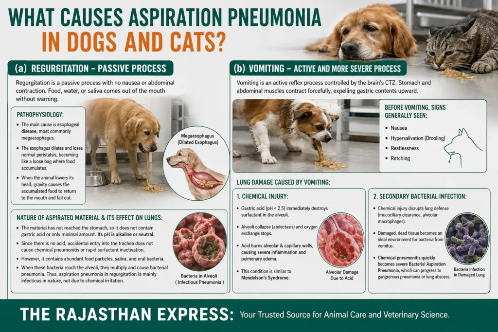
(b) Vomiting – Active and More Severe Process
Definition:
- Friends, vomiting is not something that happens easily. It is an active reflex – meaning the body deliberately and with full force does this. Its entire control is with a special part of the brain called the Chemoreceptor Trigger Zone (CTZ) . The CTZ decides when to vomit.
Before vomiting, the following are generally seen:
Vomiting does not come suddenly; there are clear signs before it, such as:
- Nausea: The animal is restless, repeatedly licking its lips.
- Hypersalivation: Excessive drooling; foam‑like saliva from the mouth.
- Restlessness: Cannot sit still, wanders around.
- Retching: Abdominal muscles strain but nothing comes out – just gagging movements.
All these are signals that vomiting is about to occur.
Lung damage caused by vomiting:
If acidic gastric contents reach the lungs, damage occurs in two phases:
1. Chemical Injury
- Destruction of Surfactant: Inside the tiny air sacs (alveoli) of our lungs, there is a very thin lubricating layer – surfactant. It is exactly like a non‑stick coating on a pan. Its job is to prevent these sacs from sticking together during exhalation and keep them open. Gastric acid, with a pH below 2.5 (very strong acid), destroys this surfactant in an instant.
- Alveolar collapse (Atelectasis): As soon as surfactant is destroyed, all the alveoli stick together during exhalation and cannot reopen no matter how hard you try. This is called atelectasis. It is exactly like a wet plastic bag sticking from the inside that you cannot inflate. Result? Breathing becomes difficult, and oxygen supply to the body stops.
- Burning of walls and Inflammation: This strong acid not only destroys surfactant but also directly burns the delicate walls of the alveoli and blood capillaries. This burn triggers a severe inflammatory response. Lung tissue swells rapidly and fills with fluid (pulmonary edema). This entire condition is considered similar to Mendelson’s Syndrome in humans – a very serious and life‑threatening chemical burn.
2. Secondary Bacterial Infection
The damage caused by chemical injury completely disrupts the local defense system of the lungs, such as mucociliary clearance and alveolar macrophages. Now, the bacteria that came with the vomit find this environment like paradise – warm, moist, full of dead tissue – a perfect breeding ground. They multiply rapidly and cause severe bacterial aspiration pneumonia. If this infection worsens, it can become gangrenous pneumonia (lung tissue starts decaying) or lung abscess (a pus‑filled nodule inside the lungs).
Major Risk Factors for Aspiration Pneumonia in Dogs and Cats – these conditions greatly increase the risk of aspiration
(a) Pharyngeal Abnormalities
- Cricopharyngeal Motor Dysfunction: The name is big, but the function is easy to understand. There is a small muscle (cricopharyngeus muscle) at the lower part of the throat. When the animal swallows, this muscle opens so that food can enter the esophagus. Sometimes this muscle does not open on time or does not function properly. Then what happens? As the animal tries to swallow, the food is pushed upward instead of down into the esophagus and can fall directly into the trachea. In this condition, you will see the animal repeatedly trying to swallow empty, coughing, and sometimes food coming out of the nose. Meaning, the coordination of swallowing is disturbed, creating a risk of aspiration.
(b) Esophageal Diseases
- Megaesophagus: The esophageal muscles stop working. The esophagus dilates into a loose bag, and food accumulates there. As soon as the dog lowers its head, due to gravity, all the accumulated food passively (regurgitation) comes back into the mouth and can easily enter the trachea. This is one of the biggest causes of aspiration pneumonia.
- Gastroesophageal Reflux Disease (GERD): In this condition, stomach acid and enzymes repeatedly come back into the esophagus, causing irritation and inflammation. Sometimes this acid reaches the throat, especially when the animal is sleeping or unconscious. This acid then descends into the lungs with breath, causing intense irritation and infection.
- Esophageal Obstruction: If a bone piece, toy, or tumor blocks the esophagus, saliva and food accumulate above it and can enter the lungs.
(c) Debilitated State
- When an animal is very sick – such as kidney failure, cancer, or any long‑term serious illness – its entire body becomes very weak. In this weakness, its normal swallowing reflex either becomes very slow or is lost. In such a condition, even saliva and a little food in the mouth cannot be swallowed properly, and everything goes straight into the trachea. Not only that, if the animal is lying down all the time, saliva and secretions can accumulate in the throat and pose a risk of entering the lungs.
(d) Brachycephalic Breeds – such as Pugs, Bulldogs
Scientific reason – Increased Negative Pressure:
These adorable‑looking pugs and bulldogs have a major physical problem. Their nose, throat, and trachea are naturally very narrow. When they breathe, they have to exert a lot of force to draw air through this tight passage. Due to this force, a very strong suction or pull (negative pressure) is created inside their chest. This strong suction works exactly like you sucking a cold drink through a straw with force. This pull forcefully draws stomach contents (food and acid) upward to the throat. And from there, it easily enters the lungs with breathing. This is why aspiration pneumonia due to reflux is very common in these breeds, even without vomiting.
(e) Anesthesia/Deep Sedation – the biggest danger
This danger arises when we put animals under anesthesia for surgery or any examination. Anesthetic drugs simultaneously eliminate two very important safety shields of the body:
- The throat’s safety guard shuts down: Our throat (larynx) has an amazing reflex. As soon as any particle of water or food accidentally reaches the opening of the trachea, the trachea’s flap closes immediately and a strong cough occurs. This is the laryngeal reflex. Anesthetic drugs eliminate this reflex. Now if vomiting occurs or saliva drips, it will go straight into the lungs without any hindrance.
- The stomach’s gate becomes loose: There is a tight valve or gate between the stomach and the esophagus, called the Lower Esophageal Sphincter (LES) . Its job is to prevent stomach contents from coming up. Anesthetic drugs completely relax this gate. Now if there is anything in the stomach, it can easily come up to the throat and then flow into the unprotected trachea.
That is why the most important preventive measure is – Pre‑Anesthetic Fasting:
This is not an old custom, but a solid rule based on science. Food is completely stopped about 8‑12 hours before surgery and water about 2‑4 hours before. This keeps the stomach completely empty. Now if the gate becomes loose under anesthesia, there is nothing in the stomach to go to the lungs. This small step can save your pet’s life.
Major Causes of Aspiration Pneumonia in Large Animals (Cattle, Buffaloes, Sheep, and Goats)
(a) Improper drenching technique
- The most common cause of aspiration pneumonia in large animals is improper oral drenching of medicine or liquid.
- When we give liquid medicine by drench or bottle/syringe, it is very important that we do not pour faster than the animal’s swallowing speed. Scientifically, the larynx of ruminants is structured such that they cannot breathe and swallow at the same time. During swallowing, the epiglottis closes the trachea. However, if the animal is stressed, coughing, bellowing, or if we have pulled its tongue and raised its head too high, this coordination breaks down. Then the medicine falls directly onto the open larynx and enters the trachea.
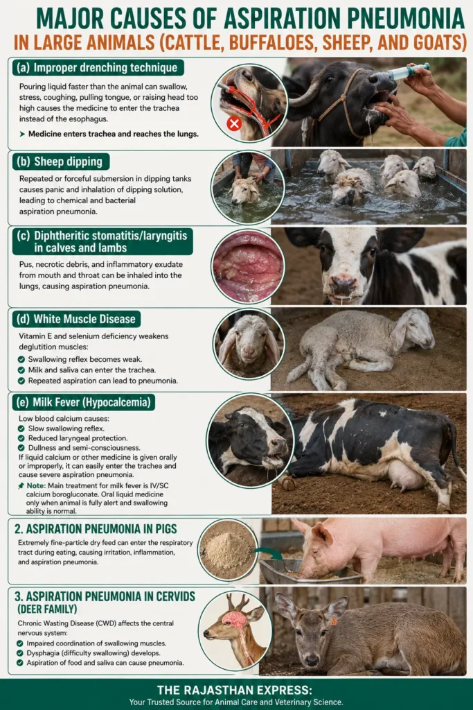
(b) Sheep dipping
- When sheep are dipped in dipping tanks for parasite control, if they are repeatedly or forcibly submerged, they may inhale the dipping solution in panic. This can cause both chemical and bacterial aspiration pneumonia.
(c) Diphtheritic stomatitis/laryngitis in calves and lambs
- If calves or lambs develop diphtheritic stomatitis or laryngitis, the pus, necrotic debris, and inflammatory exudate formed in the mouth and throat can enter the lungs with breathing and cause aspiration pneumonia.
(d) White Muscle Disease
Nutritional myopathy caused by vitamin E and selenium deficiency in lambs (White Muscle Disease) weakens the deglutition muscles (swallowing muscles). As a result:
- The swallowing reflex becomes weak.
- Milk and saliva can enter the trachea.
- Repeated aspiration can lead to pneumonia.
(e) Milk Fever (Hypocalcemia)
In milk fever, the blood calcium level drops excessively, causing:
- The swallowing reflex to become slow.
- The protective ability of the larynx to decrease.
- The animal to become dull and semi‑conscious.
- If such an animal is given liquid calcium or other medicine orally or with improper technique, it can easily enter the trachea and cause severe aspiration pneumonia.
Note: In practical veterinary medicine, the main treatment for milk fever is intravenous or subcutaneous administration of calcium borogluconate. Oral liquid medicine should only be given when the animal is fully alert and its swallowing ability is normal.
2. Aspiration Pneumonia in Pigs
- If pigs are fed extremely fine‑particle dry feed, microscopic particles can enter the respiratory tract during eating. Over time, this can cause irritation, inflammation, and aspiration pneumonia.
3. Aspiration Pneumonia in Cervids (Deer Family)
Chronic Wasting Disease (CWD) is a prion‑induced neurological disease. In this disease:
- The central nervous system is affected.
- Coordination of swallowing muscles is impaired.
- Dysphagia (difficulty swallowing) develops.
- As a result, aspiration of food and saliva can cause pneumonia.
Microbiology of Aspiration Pneumonia in Dogs – Which Bacteria Cause the Infection?
Friends, when food, water, or vomited acid goes into the lungs, it does not go alone. It carries with it a lot of bacteria from the mouth, throat, and stomach. And this is where the real game of infection begins.
The most important fact is that aspiration pneumonia in dogs is not caused by a single bacterium. Instead, it is usually a mixed bacterial infection (Mixed Infection), in which multiple bacterial species work together to infect the lungs. These bacteria are divided into two teams:
- Anaerobic bacteria (without oxygen): These organisms survive without oxygen.
- Aerobic bacteria (oxygen‑loving): These need oxygen to survive.
Now let us know these two teams in a little more detail.
1. Anaerobic Bacteria – The Hidden Cause of Tissue Necrosis and Gangrene
When lung tissue is burnt by acid or food gets lodged, the oxygen supply there decreases. In such an environment, these anaerobic bacteria multiply very rapidly and start decaying the dead tissue (flesh). That is why they can also be called decay‑causing bacteria.
Main names:
- Bacteroides
- Fusobacterium
- Prevotella
These are the same bacteria that cause foul‑smelling decay like gangrene.
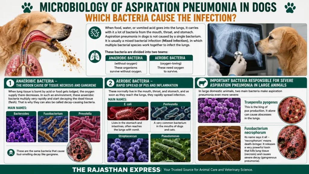
2. Aerobic Bacteria – Rapid Spread of Pus and Inflammation
These normally live in the mouth, throat, and stomach, and as soon as they reach the lungs, they rapidly spread infection.
Main names:
- E. coli: Lives in the stomach and intestines, often reaches the lungs with vomit.
- Pasteurella: A very common bacterium in the mouths of dogs and cats.
- Streptococcus
- Pseudomonas
Important Bacteria Responsible for Severe Aspiration Pneumonia in Large Animals
In large domestic animals, two main bacteria make aspiration pneumonia even more severe:
- Trueperella pyogenes: This is the king of pus production. It alone can cause abscesses in the lungs.
- Fusobacterium necrophorum: Its name says it all – ‘necrophorum’ means death‑bringer. It releases a very powerful toxin that kills lung tissue (necrosis) and causes severe decay (gangrenous pneumonia).
Symptoms of Aspiration Pneumonia in Dogs – How to Recognize the Disease Early
The symptoms of aspiration pneumonia in dogs depend entirely on how much and what type of material—such as food, water, milk, stomach acid, or oily substances—has entered the lungs.
If a large amount of acidic gastric contents, liquid, or oily material suddenly enters the lungs, aspiration pneumonia develops acutely, and the dog’s condition may deteriorate within a few hours.
On the other hand, if there is chronic regurgitation from the esophagus, where small amounts of food repeatedly enter the lungs over time, the disease develops gradually. In such cases, dogs often show chronic coughing, progressive weakness, and recurrent respiratory problems.
Let us understand the signs of aspiration pneumonia in dogs one by one in simple language.
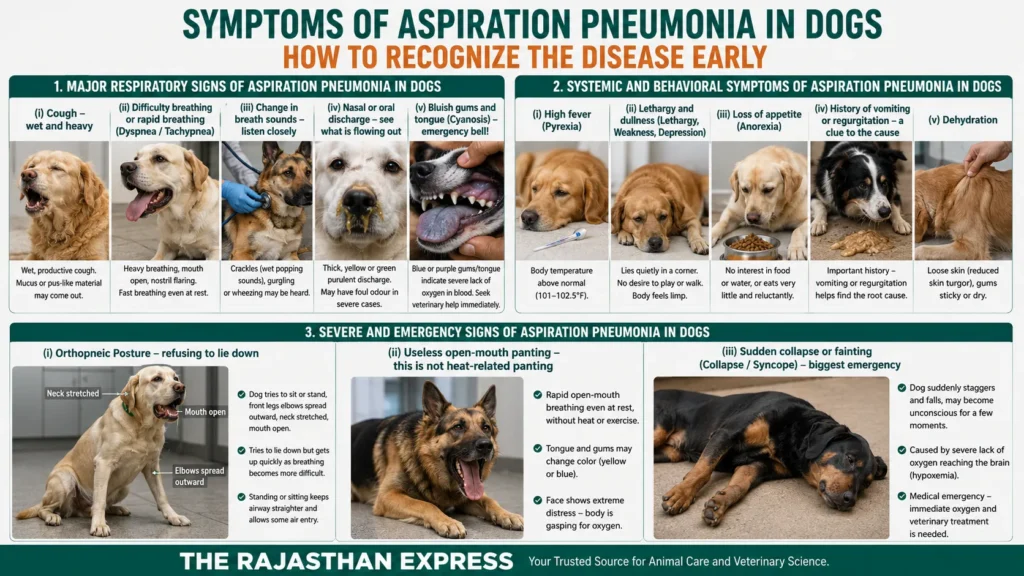
1. Major Respiratory Signs of Aspiration Pneumonia in Dogs
These are usually the first signs of aspiration pneumonia in dogs because lung damage directly affects breathing.
(i) Cough – wet and heavy
How does it feel?
- This is usually a wet/productive cough. It sounds like something is stuck in the throat and the dog is trying to expel it. Sometimes, sticky mucus or pus‑like material may also come out.
Why does this happen?
- Think about it – when foreign material and bacteria enter the bronchi and alveoli, the body’s cleaning system becomes immediately active. It shouts – “Get this out!” This is the cough reflex. Due to inflammation, excessive mucus and pus form inside the lungs, which the body tries to expel through coughing.
Dry cough can also occur:
- If acid has just gone in and only chemical pneumonitis has occurred, a dry cough may appear initially. But as soon as the infection sets in, it turns into a wet cough.
(ii) Difficulty breathing or rapid breathing (Dyspnea / Tachypnea) – gasping for air
- Dyspnea – difficulty breathing: You will see the dog breathing heavily, taking breaths with its mouth open, and the nostrils flaring repeatedly (nasal flaring). It feels like its breath is exhausted.
- Tachypnea – rapid breathing rate: The dog is sitting calmly, not moving at all, yet its breaths are very fast, as if it has just run a race.
What does science say?
- In pneumonia, most of the lungs become damaged. Either the air sacs (alveoli) are filled with pus, or they have collapsed and closed due to acid irritation (atelectasis). The result? Severe hypoxia (oxygen deficiency) in the blood, and carbon dioxide may increase. The part of the brain that controls breathing (respiratory centre) sees this crisis and sends an order – “Breathe faster, otherwise there will be no oxygen!”
(iii) Change in breath sounds – listen closely
- If you listen with a stethoscope, or sometimes even without it, strange sounds come during breathing. Like crackles – the sound is exactly like slowly tearing wet paper or plastic, or sometimes a gurgling sound.
- Sometimes wheezing (a whistling sound) can also be heard.
Why these sounds?
- Crackles are produced when air tries to pass through fluid or pus inside. Air bubbles bursting create this sound. Wheezing occurs when the airways become narrowed (bronchoconstriction) or swollen, making the passage for air very tight.
(iv) Nasal or oral discharge – see what is flowing out
- Sometimes thick, yellow or green purulent discharge comes from the nose. Similar sticky material may also come out with coughing.
- Serious warning: If the disease has progressed very far and lung tissue has died and started decaying (gangrenous pneumonia), this discharge will have a very strong, fetid (foul) odour of rotting matter. This smell is proof that dead tissue (necrotic tissue) and dangerous anaerobic bacteria (which thrive without oxygen) are present.
(v) Bluish gums and tongue (Cyanosis) – emergency bell!
- This is the most dangerous and emergency symptom. If you see the dog’s gums, tongue, or the pink part inside the eyes turning blue or purple, understand that the condition is very serious.
- Scientific reason: The red colour of blood is due to oxygen. When the oxygen level in the blood becomes very low (severe hypoxemia), deoxygenated hemoglobin starts showing its true dark blue colour. This means the lungs have almost failed and are unable to supply oxygen to the blood. If you see this, consult a doctor immediately.
2. Systemic and Behavioral Symptoms of Aspiration Pneumonia in Dogs
Apart from respiration, the effect of this disease is clearly visible on the entire body.
(i) High fever (Pyrexia)
Body temperature goes above normal (101‑102.5°F). The stomach feels clearly hot.
Why does fever occur? When bacteria enter and attack and there is inflammation in the tissue, our body’s macrophages release some special chemical messengers called pyrogens (such as IL‑1, IL‑6). These messengers go to the brain’s temperature control centre (hypothalamus) and forcefully raise the body’s thermostat. Fever is actually the body’s defence, but if it crosses 104°F, it can be dangerous in itself.
(ii) Lethargy and dullness (Lethargy, Weakness, Depression)
- The dog will lie quietly in a corner. No desire to play or walk. If you tell it to get up, it stands with great difficulty, the body feels completely limp.
(iii) Loss of appetite (Anorexia)
- Keep food and water in front – it won’t even turn its face to it, or will eat very little and reluctantly.
(iv) History of vomiting or regurgitation – a clue that tells the root of the disease
- This is not a symptom, but a very important question that the doctor will definitely ask you. “Did the dog vomit recently? Or did food come out of the mouth without effort?”
Why is this important? This information immediately tells the doctor where the disease started. Food coming out without effort (regurgitation) indicates esophageal disease, while forceful vomiting means that stomach acid has entered the lungs, causing severe chemical injury.
(v) Dehydration
- You will see that the dog’s skin has become loose (reduced skin turgor), and the gums feel sticky or dry to the touch.
3. Severe and Emergency Signs of Aspiration Pneumonia in Dogs
These are the symptoms that indicate the disease has gone beyond the limit of tolerance. The dog’s life is directly in danger. It is very important to recognise them.
(i) Orthopneic Posture – refusing to lie down
What does this look like?
- The dog deliberately tries to sit or stand. You will see its front legs’ elbows spread outward, neck stretched, and mouth open. It will try to lie down repeatedly, but as soon as it lies down, breathing becomes even more difficult and it quickly gets up and sits. It is completely afraid to lie down.
Why?
- When lying down, the abdominal organs (liver, stomach) press on the diaphragm (the main breathing muscle). When the lungs are already filled with pus and inflammation, this pressure compresses them further. Therefore, by standing or sitting, stretching its neck, the dog keeps its airway as straight as possible, so that some air can enter. This is the body’s forced posture.
(ii) Useless open‑mouth panting – this is not heat‑related panting
Note: Dogs pant in heat or after exercise – this is normal. But here the situation is different. If the dog, without any heat or exercise, just sitting, is continuously breathing rapidly with its mouth open, this is not fun, but a sign of respiratory distress.
How to recognise?
- In this panting, the colour of the tongue and gums may change (yellow or blue), and the face will show extreme distress. This means that breathing through the nose is not enough – the body is gasping for oxygen and has to draw air in every way possible.
(iii) Sudden collapse or fainting (Collapse / Syncope) – biggest emergency
What is this? The dog suddenly staggers and falls while walking or sitting, and may become unconscious for a few moments. Its body becomes completely limp.
Why does this happen?
- This occurs when the oxygen deficiency in the blood (hypoxemia) becomes so severe that oxygen stops reaching the brain. For the brain, oxygen is everything – without oxygen it cannot function properly even for a moment. As soon as the brain did not get enough oxygen, it lost balance and consciousness.
- This is not ordinary fainting, but a medical emergency. If this happens, understand that the lungs have almost completely failed and immediate oxygen and treatment are needed. Without any delay, immediately take the dog to the hospital and get treatment from a veterinarian.
Diagnosis of Aspiration Pneumonia in Dogs
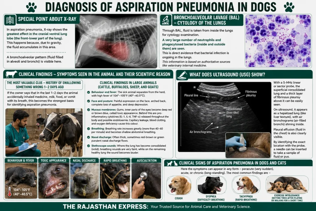
Special point about X‑ray:
- In aspiration pneumonia, X‑ray shows the greatest effect in the cranial ventral lung lobe (the front‑lower part of the lung). This happens because, due to gravity, the fluid accumulates in this area. A bronchoalveolar pattern (fluid filled in alveoli and bronchi) is visible here.
Bronchoalveolar Lavage (BAL) – Cytology of the Lungs
- Through BAL, fluid is taken from inside the lungs for cytology examination. A very large number of neutrophils and phagocytosed bacteria (inside and outside them) are seen. This is direct evidence that bacterial infection is ongoing in the lungs. This information is based on authoritative sources like veterinary internal medicine.
Clinical Findings – Symptoms seen in the animal and their scientific reason
The most valuable clue – history of swallowing something wrong 1‑2 days ago
If the owner says that in the last 1‑2 days the animal accidentally inhaled medicine, milk, food, or vomit with its breath, this becomes the strongest basis for identifying aspiration pneumonia.
Clinical Findings in Large Animals (Cattle, Buffaloes, Sheep, and Goats)
- Behaviour and fever: The sick animal separates from the herd, with high fever of 104°‑105°F (40°‑40.5°C) .
- Face and posture: Painful expression on the face, arched back, complete loss of appetite, and deep depression.
- Mucous membranes: Gums, inner parts of the eyes become deep red or brown‑blue, called toxic appearance. Behind this are pro‑inflammatory cytokines (IL‑1, IL‑6, TNF‑α) released throughout the body and possible endotoxemia. Capillary leakage, blood clotting, and oxygen deficiency cause this colour.
- Breathing: Breathing rate increases greatly (more than 40‑60 per minute) and becomes shallow abdominal breathing.
- Nasal discharge: Often thick, sometimes red‑brown or green purulent nasal discharge flows.
- Stethoscope sounds: Where the lung has become consolidated (solid), breathing sounds are very faint, while on the remaining healthy lung the sound becomes louder.
What does ultrasound (USG) show?
With a 5‑MHz linear or sector probe, the superficial consolidated lung and a thick layer of fibrinous pleurisy above it can be easily seen. On ultrasound, it appears as a hepatized lung (like liver texture), with air bronchograms (air‑filled bronchi) shining inside. Pleural effusion (fluid in the chest) is also clearly visible. By identifying the exact location with the probe, a needle can be inserted to take a sample of fluid or pus.
Clinical Signs of Aspiration Pneumonia in Dogs and Cats
- Here the symptoms can appear in any form – peracute (very sudden), acute, or chronic (long‑standing). The most common findings are – cough, dyspnea (difficulty breathing), tachypnea (rapid breathing), and exercise intolerance (getting tired after playing or walking for a short time).
Prevention and Treatment of Aspiration Pneumonia in Dogs (Aspiration Pneumonia Dogs Treatment)
Friends, this disease can become severe very quickly, so we must either prevent it from occurring, and if it does occur, start the right treatment without delay. Let us understand both one by one.
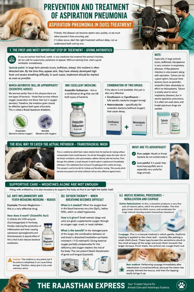
1. The first and most important step of treatment – Giving antibiotics
- If you are certain that food, vomit, or any medicine has entered the animal’s trachea, do not wait for pneumonia symptoms to appear. Without wasting time, start a good antibiotic immediately.
- Special point: In large farm animals (cows, buffaloes, sheep), this incident is often detected late. By the time they appear sick, they have already developed high fever and severe breathing difficulty. In such cases, treatment should be started as soon as possible.
Which antibiotic will be appropriate? (Scientific advice)
We learned earlier that in this disease there are two types of bacteria – those that survive without oxygen (anaerobic) and those that live in oxygen (aerobic). Therefore, the medicine given should be effective against both types of bacteria. This is called a Broad‑Spectrum Antibiotic.
- One excellent medicine: Ampicillin‑Sulbactam – this is a combination drug that can kill both types of bacteria.
- Combination of two drugs: If the above is not available, this pair is also very effective:
- Enrofloxacin or Ciprofloxacin – kills aerobic bacteria (oxygen‑loving).
- Metronidazole – specifically for anaerobic bacteria (without oxygen) that cause decay.
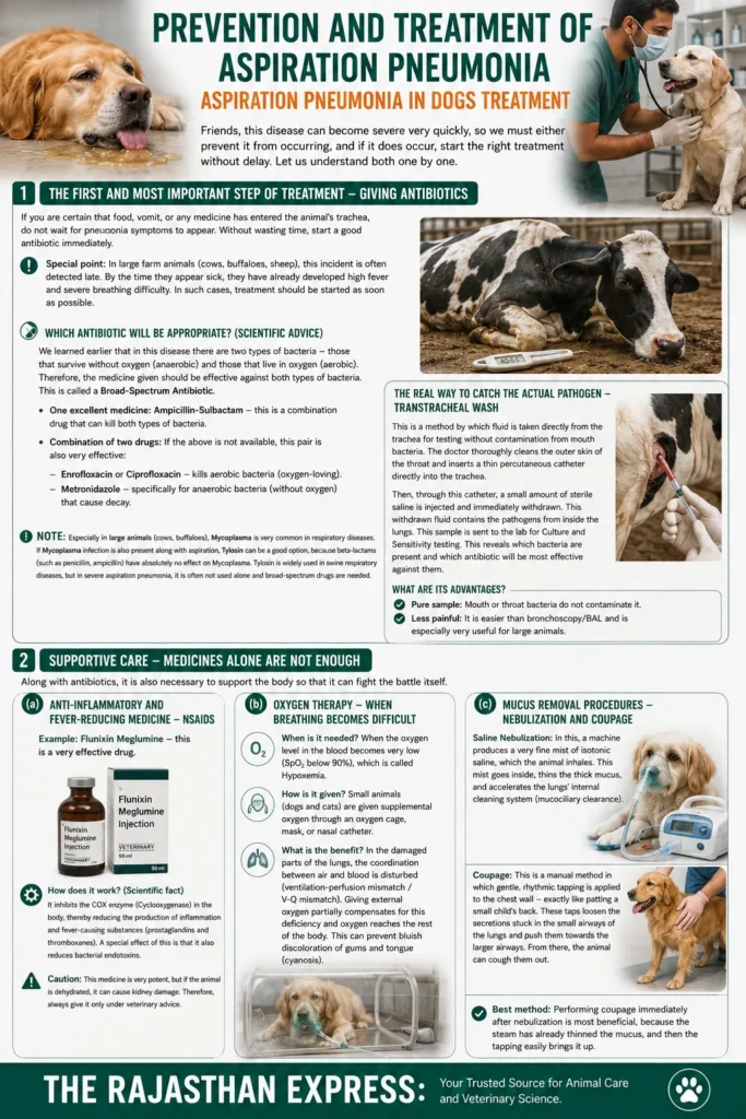
Especially in large animals (cows, buffaloes), Mycoplasma is very common in respiratory diseases. If Mycoplasma infection is also present along with aspiration, Tylosin can be a good option, because beta‑lactams (such as penicillin, ampicillin) have absolutely no effect on Mycoplasma. Tylosin is widely used in swine respiratory diseases, but in severe aspiration pneumonia, it is often not used alone and broad‑spectrum drugs are needed.
THE RAJASTHAN EXPRESS
The real way to catch the actual pathogen – Transtracheal Wash
This is a method by which fluid is taken directly from the trachea for testing without contamination from mouth bacteria. The doctor thoroughly cleans the outer skin of the throat and inserts a thin percutaneous catheter directly into the trachea. Then, through this catheter, a small amount of sterile saline is injected and immediately withdrawn. This withdrawn fluid contains the pathogens from inside the lungs. This sample is sent to the lab for Culture and Sensitivity testing. This reveals which bacteria are present and which antibiotic will be most effective against them.
What are its advantages?
- Pure sample: Mouth or throat bacteria do not contaminate it.
- Less painful: It is easier than bronchoscopy/BAL and is especially very useful for large animals.
Supportive Care – Medicines alone are not enough
Along with antibiotics, it is also necessary to support the body so that it can fight the battle itself.
(a) Anti‑inflammatory and fever‑reducing medicine – NSAIDs
- Example: Flunixin Meglumine – this is a very effective drug.
- How does it work? (Scientific fact) It inhibits the COX enzyme (Cyclooxygenase) in the body, thereby reducing the production of inflammation and fever‑causing substances (prostaglandins and thromboxanes). A special effect of this is that it also reduces bacterial endotoxins.
- Caution: This medicine is very potent, but if the animal is dehydrated, it can cause kidney damage. Therefore, always give it only under veterinary advice.
(b) Oxygen Therapy – when breathing becomes difficult
- When is it needed? When the oxygen level in the blood becomes very low (SpO₂ below 90%), which is called Hypoxemia.
- How is it given? Small animals (dogs and cats) are given supplemental oxygen through an oxygen cage, mask, or nasal catheter.
- What is the benefit? In the damaged parts of the lungs, the coordination between air and blood is disturbed (ventilation‑perfusion mismatch / V‑Q mismatch). Giving external oxygen partially compensates for this deficiency and oxygen reaches the rest of the body. This can prevent bluish discoloration of gums and tongue (cyanosis).
(c) Mucus removal procedures – Nebulization and Coupage
- Saline Nebulization: In this, a machine produces a very fine mist of isotonic saline, which the animal inhales. This mist goes inside, thins the thick mucus, and accelerates the lungs’ internal cleaning system (mucociliary clearance).
- Coupage: This is a manual method in which gentle, rhythmic tapping is applied to the chest wall – exactly like patting a small child’s back. These taps loosen the secretions stuck in the small airways of the lungs and push them towards the larger airways. From there, the animal can cough them out.
- Best method: Performing coupage immediately after nebulization is most beneficial, because the steam has already thinned the mucus, and then the tapping easily brings it up.
Prevention is the best treatment (Prognosis & Prevention of Aspiration Pneumonia in Dogs)
Friends, despite so many treatments, the prognosis for aspiration pneumonia remains very poor. Meaning, even after full effort, it may be difficult to save the animal, especially when acid burns or gangrene have occurred.
Therefore, all effort should be focused on prevention. For example:
- Caution while drenching: Do not raise the head too high, do not pull the tongue, and do not pour faster than the animal’s swallowing speed.
- Fasting before anesthesia: Stopping food 8‑12 hours before surgery and water 2‑4 hours before is the easiest and life‑saving step.
- Special care for brachycephalic breeds (Pugs, Bulldogs): Feed them in small portions with their head facing upward, and protect them from excessive heat/stress.
- Always feed dogs with megaesophagus in an upright position: Use a special “Bailey Chair” so that gravity helps food reach the stomach.
Remember, in this disease, “prevention is better than cure” – this saying is 100% true. A small precaution can save your pet companion’s life.
Aspiration pneumonia is one of the most serious respiratory emergencies in veterinary medicine. Recognizing the first signs of aspiration pneumonia in dogs, seeking immediate veterinary care, and preventing aspiration through proper feeding, anesthesia protocols, and management of underlying diseases are the most effective ways to improve recovery and survival.
THE RAJASTHAN EXPRESS
Source By : THE MERCK VETERINARY MANUAL ELEVENTH EDITION
People Also Ask
What is aspiration pneumonia in dogs?
What causes aspiration pneumonia in dogs?
- Ingesta: Food or water going down the wrong pipe during swallowing.
- Regurgitation: Passive return of food from the esophagus (common in megaesophagus).
- Vomiting: Forceful expulsion of acidic stomach contents into the lungs.
What are the signs of aspiration pneumonia in dogs?
- Wet/productive cough – often with mucus or pus.
- Difficulty breathing (Dyspnea) – laboured breathing, open mouth, nasal flaring.
- Rapid breathing (Tachypnea) – fast breathing even at rest.
- Abnormal breath sounds – crackles (rales) or wheezing.
- Nasal discharge – thick, yellow/green purulent discharge.
- Fever, lethargy, loss of appetite, and dehydration.
- Cyanosis – blue/purple gums and tongue (emergency sign).
What are the first signs of aspiration pneumonia in dogs?
- Coughing after eating or drinking
- Gagging or retching
- Laboured or rapid breathing
- Lethargy and weakness
- Loss of appetite
What are the stages of aspiration pneumonia in dogs?
- Initial Aspiration: Foreign material enters the trachea and lungs.
- Chemical Pneumonitis: If acidic gastric contents are aspirated, they cause rapid destruction of surfactant, alveolar collapse, and severe inflammation (similar to Mendelson’s Syndrome).
- Bacterial Infection: Bacteria from the aspirated material multiply in the damaged lung tissue, causing bacterial pneumonia.
- Severe Pneumonia: Widespread infection with pus formation, consolidation, and respiratory distress.
- Gangrenous Pneumonia or Lung Abscess: In severe cases, lung tissue necrosis, gangrene, or abscess formation occurs.
What are the end stages of aspiration pneumonia in dogs?
- Severe respiratory distress – extreme difficulty breathing, open-mouth panting.
- Cyanosis – blue/purple gums and tongue due to severe oxygen deficiency.
- Collapse or syncope – sudden fainting due to severe hypoxemia.
- Respiratory failure – the lungs can no longer oxygenate the blood.
- Without immediate veterinary intervention, this can be fatal.
What is the survival rate of aspiration pneumonia in dogs?
- Timely diagnosis and treatment: Early intervention significantly improves outcomes.
- Severity of aspiration: Small volume, non-acidic aspiration has better prognosis than large volume, acidic aspiration.
- Presence of gangrene or abscesses: These complications lower the survival rate.
- Underlying conditions: Megaesophagus, brachycephalic syndrome, or immunosuppression worsen prognosis.
How to treat aspiration pneumonia in dogs?
- Broad-spectrum antibiotics: Ampicillin-Sulbactam, or Enrofloxacin + Metronidazole combination to target both aerobic and anaerobic bacteria.
- Oxygen therapy: For dogs with hypoxemia (SpO₂ below 90%).
- NSAIDs: Flunixin Meglumine to reduce fever and inflammation (use with caution if dehydrated).
- Nebulization and Coupage: To thin mucus and help expel secretions.
- Fluid therapy: To correct dehydration.
- Transtracheal wash and culture: To identify the exact bacteria and choose the most effective antibiotic.
- Severe cases may require hospitalization and intensive care.
What is the recovery time for aspiration pneumonia in dogs?
- Severity of the condition: Mild cases may recover in 1-2 weeks with treatment.
- Moderate cases: May take 3-4 weeks of antibiotic therapy and supportive care.
- Severe cases: With complications like abscesses or gangrene, recovery can take several weeks to months.
- Underlying conditions: Dogs with megaesophagus or brachycephalic syndrome may take longer or have recurrent episodes.
Are brachycephalic breeds at higher risk for aspiration pneumonia?
What is the difference between regurgitation and vomiting in dogs?
- Regurgitation is a passive process where food, water, or saliva comes out of the mouth without warning, effort, or nausea. It is caused by esophageal diseases like megaesophagus. Aspirated material from regurgitation is neutral/alkaline and causes infectious (bacterial) pneumonia.
- Vomiting is an active reflex controlled by the brain’s Chemoreceptor Trigger Zone (CTZ). It involves forceful contractions of the stomach and abdominal muscles, preceded by signs like nausea, drooling, and retching. Aspirated material from vomiting is acidic and causes chemical pneumonitis followed by bacterial infection.
How can I prevent aspiration pneumonia in my dog?
- Proper feeding technique: Never force-feed a weak or sedated dog. Feed puppies on their stomach, not on their back.
- Fasting before anesthesia: Withhold food for 8-12 hours and water for 2-4 hours before surgery.
- Special care for brachycephalic breeds: Feed in small portions with head elevated; avoid excessive heat and stress.
- Megaesophagus management: Feed dogs in an upright position using a Bailey Chair so gravity helps food reach the stomach.
- Caution while drenching: Do not raise the head too high or pull the tongue; pour slowly.


