Caprine Arthritis/Encephalitis (CAE) in Goats and Sheep
Q: Can goats recover from Caprine Arthritis Encephalitis?
A: No – CAE has no cure. Once infected, goats remain lifelong carriers. The virus hides in their immune cells, causing irreversible joint damage, udder fibrosis, and 19-34% milk loss. Early detection through blood tests (AGID/ELISA) is critical to isolate carriers and save your herd.”**
THE RAJASTHAN EXPRESS
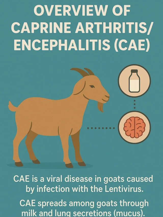
Overview of Caprine Arthritis/Encephalitis (CAE)
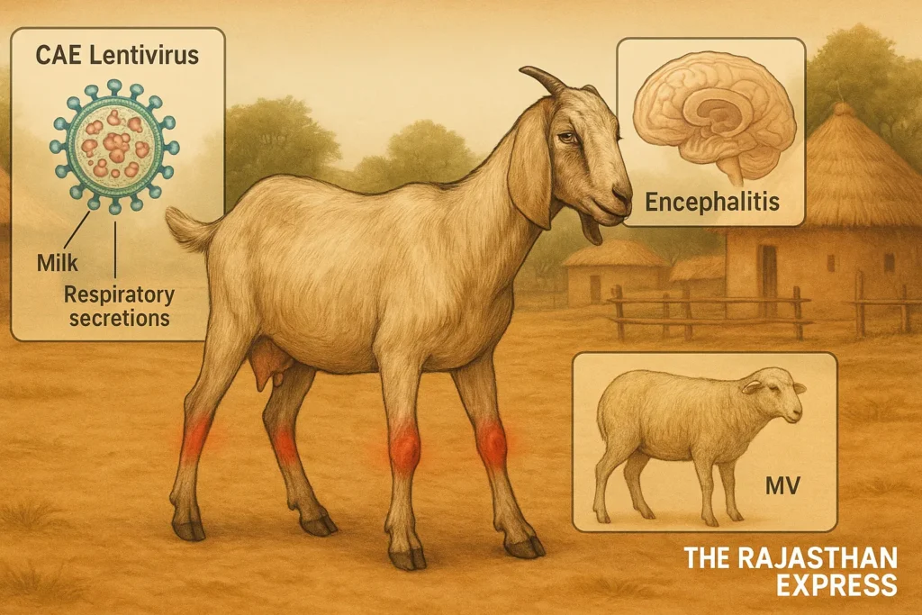
Caprine Arthritis-Encephalitis (CAE) is a viral disease in goats caused by infection with the Lentivirus. CAE spreads among goats through milk and lung secretions (mucus). It is a chronic condition, meaning it infects the animal for a long duration.
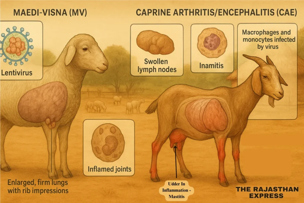
A similar disease occurs in sheep, known as Maedi-Visna (MV). Both diseases are classified under the Lentivirus group because comparison of the nucleotide sequences of the MV virus (MVV) and CAE virus (CAEV) shows they are closely related. The viruses share significant similarities. Maedi-Visna is also referred to as Ovine Progressive Pneumonia (OPP).

Caprine Arthritis Encephalitis (CAE) in Goats
| Alternate Names | CAE, Small Ruminant Lentivirus (SRLV) infection, Ovine Progressive Pneumonia (sheep; MVV), Maedi-Visna (sheep) |
|---|---|
| Definition | Chronic, lifelong lentiviral infection in goats characterized by multisystemic inflammation, including arthritis, encephalitis, mastitis, pneumonia, and wasting. No cure exists. |
| Epidemiology & Prevalence | Widespread globally; prevalence in some herds may exceed 80%. In the U.S., ~73% of herds had at least one seropositive goat; California dairies show ~53% individual seroprevalence. |
| Transmission | Primarily via ingestion of infected colostrum/milk. Less commonly through respiratory secretions, blood, equipment, and in utero. |
| Pathogenesis | The virus hides in monocytes/macrophages and persists indefinitely. Inflammatory lesions are common in joints, brain, lungs, udder, and occasionally kidneys and heart. |
| Clinical Presentations |
|
| Diagnosis |
|
| Treatment & Management | No antiviral therapy exists. Supportive care only. Culling or permanent isolation of positives is recommended. |
| Prevention & Control |
|
| Economic Impact | Infected goats suffer decreased milk yield, breeding value loss, udder fibrosis, and diminished productivity. |
| Sources: USDA-ARS; Cooperative Goat Extensions; MSD Vet Manual; Minnesota BAH OPP/CAE Program; CFSH-Iowa State; The Rajasthan Express | |
Meaning of “Maedi-Visna”
The term “Maedi-Visna” comes from the Icelandic language and represents two types of symptoms:
- “Maedi” means difficulty in breathing, which is linked to lung disease (interstitial pneumonitis).
- “Visna” means shrinking or wasting of the body, associated with swelling of the membranes surrounding the brain and central nervous system (meningoencephalitis).
Types of Meninges:
- Dura Mater
- Arachnoid
- Pia Mater
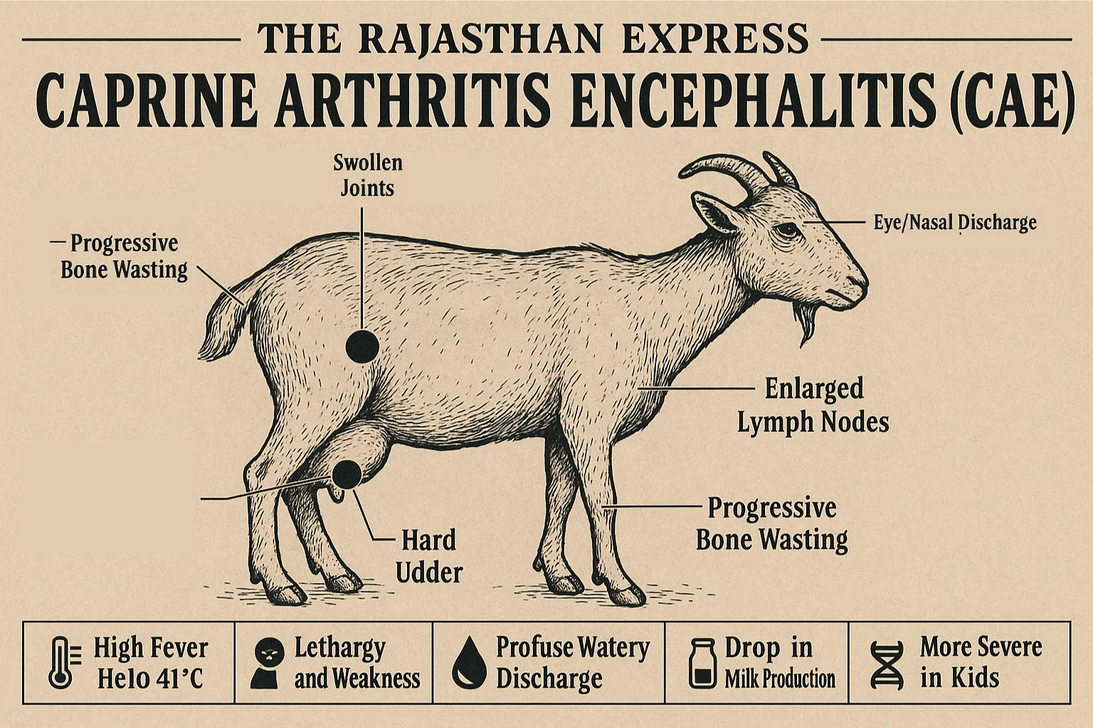
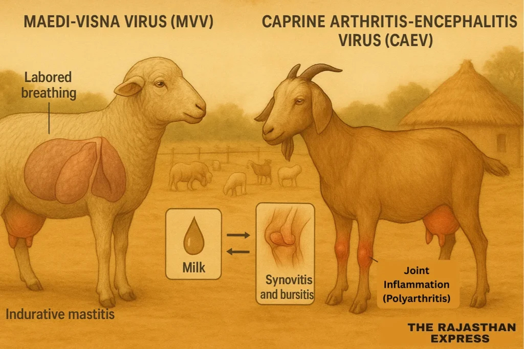
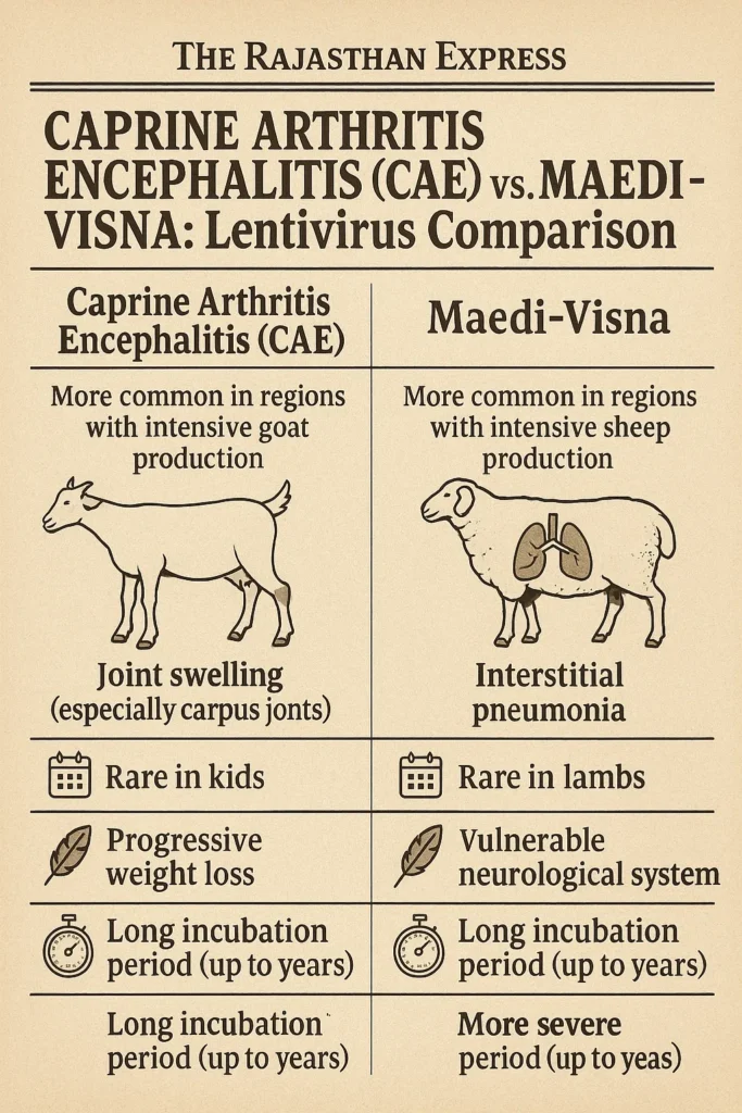
Difference Between CAE and MV in Symptoms
- MV Virus (Sheep) – Primarily causes lung disease.
- CAE Virus (Goats) – Causes joint inflammation (polyarthritis), synovitis (inflammation of the synovial membrane of the joints), and bursitis (inflammation of fluid sacs around the joints).
In young goats (2–6 months old), CAE infection can lead to brain inflammation (encephalitis). In both diseases, hard swelling of the udder (indurative mastitis) is observed.
Changes in Lungs and Other Organs
In sheep infected with MV:
- Lungs do not collapse after removal from the chest and show rib impressions.
- Lungs and lymph nodes may be 2–3 times heavier than normal due to swelling.
- Lungs become firm, greyish-brown, with evenly spread lesions.
- Udder becomes completely hardened, and associated lymph nodes may enlarge.
Cross-Species Infection
Scientific experiments have shown that:
- Sheep can be infected with CAE virus.
- Goats can be infected with MV virus.
DNA sequence analysis confirms these viruses can spread between species. However, the original source species remains unclear.
How the Virus Survives in the Body
A unique feature of CAE and MV viruses is that once an animal is infected, the virus hides in monocytes and macrophages (immune cells) for life. Most chronic diseases target these cells.
For example, tuberculosis (a bacterial disease) also increases monocyte count and forms Langerhans giant cells—typical in chronic infections.
- Monocytes – Macrophages found in the blood.
- Macrophages – Large phagocytic cells found in connective tissues.
The time between virus infection and antibody production varies. Most infected sheep and goats show no symptoms but can still spread the virus.
Global Distribution of CAE and MV Viruses
- Found in most sheep-rearing countries except Australia and New Zealand.
- CAE is mainly seen in goats from developed countries, especially European dairy breeds like Saanen, Alpine, and Toggenburg, more than in other breeds.
- Spread occurs via infected milk and respiratory secretions.
Both CAE and MV affect lungs, joints, udder, and brain, causing inflammation in these organs. Mastitis is common and reduces milk yield. Untreated mastitis can lead to fibrosis of the udder, permanently stopping milk production in the affected half.
Caprine Arthritis Encephalitis Symptoms in Goats and Sheep
Early Stage
- Often no visible symptoms, making diagnosis difficult.
- As a viral disease, antibiotics and most medicines are ineffective.
In Sheep (MVV Infection)
- Slowly progressing lung disease.
- Main symptom: lung hardening.
In Goats (CAEV Infection)
- Chronic joint swelling (polyarthritis).
- Includes synovitis and bursitis.
In Goat Kids (2–6 Months Old)
- Encephalitis (brain inflammation) is common.
- Differential diagnosis is essential to confirm the disease.
Caprine Arthritis Encephalitis Prevention
Currently, no vaccine is available for CAE.
- The only effective preventive measure is identification and isolation of infected animals.
- Although vaccination is recommended in general herd management, no vaccine for CAE exists yet.
- CAE is not zoonotic—it does not spread from goats or sheep to humans.
Import Recommendations for Breeding Goats
Veterinary authorities in importing countries should ensure:
- Animals show no signs of CAE on the day of shipment.
- Goats over 1 year old test negative within 30 days before shipment.
- Alternatively, the herd has been free from CAE for the past 3 years, with no new animals from lower health status herds introduced.
Economic Losses Caused by CAE
- Reduction in milk production.
- Permanent loss of milk from udder fibrosis.
- Unsuitability for breeding.
- Significant financial losses for farmers.
Diagnosis and Treatment Gaps
- Early-stage CAE shows minimal symptoms, making detection difficult.
- No cure: Antibiotics/medicines are ineffective against the virus.
- Udder fibrosis causes irreversible milk production loss.
Diagnosis of Caprine Arthritis/Encephalitis (CAE) in Goats and Sheep
Key Organs for Examination
The following organs are important for diagnosing caprine arthritis encephalitis in goats and sheep:
| Key Organs for Examination | |
| Organ | Diagnostic Purpose |
|---|---|
| Lungs | Detect interstitial pneumonitis (lung inflammation) |
| Brain & Spinal Cord | Identify meningoencephalitis (brain inflammation) |
| Udder | Confirm indurative mastitis (hard udder) |
| Joints & Synovium | Identify arthritis |
| Kidney | Detect vasculitis (blood vessel inflammation) |
| Key Organs for Examination | |
1. Blood Tests and Clinical Examination (Serology + Clinical Evaluation)
The simplest way to diagnose caprine arthritis encephalitis is by combining blood tests with clinical observation of symptoms.
Diagnosis considers:
- The specific testing method used.
- Similarity between the infecting virus strain and the one used in the test.
- The type of viral proteins used in the test.
2. Identification of the Virus (Agent Detection)
Virus identification is carried out in the laboratory using blood, milk, lung, or udder tissue samples from live or dead animals.
This test is highly specific, though sensitivity can vary.
Confirmation is done through immuno-labeling and electron microscopy.
3. DNA-Based Testing (PCR)
PCR (Polymerase Chain Reaction) is used to detect viral DNA.
- It is a fast and accurate technique.
- PCR results can be confirmed through cloning and sequencing.
- PCR is a standard method for identifying viruses in most viral diseases.
4. Antibody Testing (Serological Tests)
Infected goats and sheep develop specific antibodies as the infection acts like an antigen, triggering the immune system.
These antibodies can be detected through AGID (Agar Gel Immunodiffusion) and ELISA tests.
Note:
- Vaccines (if available) use inactivated or attenuated virus to act as antigens, prompting antibody formation.
- Even without vaccination, animals naturally develop antibodies against the virus.
- Other specialized methods, such as Western Blot and Radio Immunoprecipitation Tests, are available in select labs.
- Antibody detection from milk is useful for dairy goat farms.
Diagnostic Test Methods and Their Purposes
| Diagnostic Test Methods and Their Purposes | |||||||
| Method | Population Freedom from Infection | Individual Pre-Movement Testing | Eradication | Clinical Case Confirmation | Surveillance (Prevalence) | Immune Status Post-Vaccination | Agent Identification |
|---|---|---|---|---|---|---|---|
| Virus Isolation | – | – | – | + | – | – | + |
| Antigen Detection | – | – | – | + | – | – | + |
| PCR | + | + | ++ | ++ | ++ | _ | ++ |
| AGID | + | +++ | ++ | +++ | +++ | + | + |
| CFT | – | – | – | – | – | – | – |
| ELISA | +++ | +++ | +++ | + | +++ | + | + |
| VN | – | – | – | – | – | +++ | – |
| IFAT | – | + | – | – | + | – | – |
| Diagnostic Test Methods and Their Purposes | |||||||
Key:
- +++ = Highly recommended
- ++ = Recommended with limitations
- + = Limited use
- – = Not suitable
- _ = No data
1. Virus Identification
MVV and CAEV are usually not distinguished for diagnostic purposes, as both persist in the body for long periods. Detecting antibodies is generally sufficient for confirmation.
However, as antibody development takes time, recently infected animals may test negative initially.
1.1 Isolation from Live Animals
1.1.1 Maedi-Visna Virus (MVV)
- MVV DNA is found in monocytes and macrophages in the blood.
- Isolation involves culturing blood or milk cells with Sheep Choroid Plexus (SCP) cells.
Procedure:
- Collect blood sample and extract buffy coat by centrifugation (1000g, 15 min).
- Wash cells and suspend in medium at 1 million cells/ml.
- Add to SCP cell monolayer.
- Incubate at 37°C with 5% CO₂.
- After several weeks, check for cytopathic effects (CPE) such as star-shaped cells, dendrites, and syncytia.
- If unclear, confirm using immunofluorescence, immunoperoxidase, electron microscopy, or reverse transcriptase assay.
1.1.2 Caprine Arthritis Encephalitis Virus (CAEV)
- Identified in the same way as MVV.
- Initially isolated from the synovial membrane of joints.
- Peripheral blood or synovial fluid is collected.
- Goat Synovial Membrane (GSM) cells serve as indicator cells.
1.2 Isolation from Dead Animals (Post-Mortem)
- Collect lungs, synovial membrane, udder, etc., under sterile conditions.
- Cut into small pieces, place in flasks with growth medium, and incubate at 37°C with 5% CO₂.
- After several days, cells migrate out and grow.
- Prepare monolayers and test for CPE.
- Lung rinse macrophage cultures are quick to prepare and can be examined via serology, electron microscopy, and reverse transcriptase assay.
1.3 Molecular Methods
- PCR detects MV and CAE viral DNA and measures viral load.
- Confirm results with Southern blot, in situ hybridization, or sequencing.
- Real-Time PCR determines viral quantity and strain prevalence in specific regions.
- LAMP (Loop-Mediated Isothermal Amplification) is a newer, fast, and simple method for CAE detection.
PCR Challenges:
- False positives can occur due to host DNA amplification.
- Always confirm PCR products via hybridization, restriction enzyme analysis, or sequencing.
2. Serological Testing
Lentivirus infections in goats and sheep are lifelong, producing permanent antibodies.
Since MVV and CAEV antigens are highly similar, separate tests are usually unnecessary.
Common Serological Tests:
- AGID (Agar Gel Immunodiffusion) – Developed in 1973
- ELISA (Enzyme-Linked Immunosorbent Assay) – Developed in 1982
2.1 AGID Test
- Detects major viral antigens: gp135 (surface glycoprotein) and p28 (nucleocapsid protein).
- Sensitivity depends on the antigen and strain used.
- gp135-based AGID is more sensitive than p28-based AGID.
- U.S. uses WLC-1 strain for AGID testing.
Medium Composition:
- 0.7–1% agarose
- 0.05M Tris buffer (pH 7.2)
- 8% NaCl
2.2 ELISA Test
Over 30 ELISA variants detect antibodies to MVV or CAEV.
Types:
- Indirect ELISA (I-ELISA) – Uses purified virus antigen, recombinant proteins, or synthetic peptides.
- Competitive ELISA (C-ELISA) – Measures competition between test antibodies and reference antibodies.
ELISA can be applied to milk or colostrum but is less effective for detecting early infections.
2.2.1 Indirect ELISA Procedure:
- Dilute serum (1:20) and add 0.1–0.2 ml to each well.
- Incubate for 30–90 minutes.
- Wash plate and add HRP-labeled anti-ruminant Ig conjugate.
- Add chromogen, stop reaction, and read absorbance at 405 nm.
Result Interpretation:
- <30% absorption = Negative
- 30–40% = Suspicious
- ≥40% = Positive
2.2.2 Competitive ELISA Procedure:
- Add undiluted serum (0.05 ml/well).
- Incubate for 1 hour at room temperature.
- Wash, add antibody-peroxidase conjugate, and incubate for 30 min.
- Wash, add TMB chromogen, incubate for 20 min.
- Stop and read at 620–650 nm.
Result Interpretation:
- Goats: >33.2% inhibition = Positive
- Sheep: >20.9% inhibition = Positive
Source By : WOAH (World Organisation for Animal Health)
“56% of goat herds carry CAE virus – but most show no symptoms until it’s too late. Discover how to spot early warning signs (arthritis, hard udder, encephalitis), prevent transmission, and avoid devastating economic losses. Protect your livestock now.”
THE RAJASTHAN EXPRESS